Search results (5 results)
-
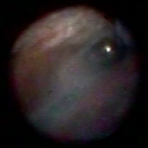 Endoscopic View: the Beginning of Anterior Hyaloid Membrane Pneumatic Dissection
Oct 2 2019 by Radwan S. Ajlan, MBBCh, FRCS(C)
Endoscopic view: the beginning of anterior hyaloid membrane pneumatic dissection.
Condition/keywords: endoscopy, hyaloid, membranes
-
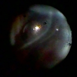 Endoscopic View - The Start of Anterior Hyaloid Membrane Pneumatic Dissection
Oct 2 2019 by Radwan S. Ajlan, MBBCh, FRCS(C)
Endoscopic view - the start of anterior hyaloid membrane pneumatic dissection.
Condition/keywords: endoscopy, hyaloid, membranes
-
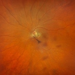 Cloquet Canal
Sep 20 2024 by Jordyn Beckman
79 year old male with wet AMD, no PVD, presents with stable cloquet canal.
Photographer: Jordyn Beckman
Imaging device: Optos California
Condition/keywords: Cloquet Canal, hyaloid membrane, punctum caecum, stilling's canal
-
 Pre Retinal Hemorrhage
Jan 11 2025 by rohan jain
Pre retinal hemorrhage
Photographer: Dr. ROHAN JAIN
Condition/keywords: Haemorrhage, hemorrhage, hyaloid membrane
-
 Advanced Proliferative Diabetic Retinopathy
Apr 9 2025 by Gustavo Uriel Fonseca Aguirre
B-mode ultrasound of a patient with long-standing poorly controlled diabetes demonstrates characteristic findings of advanced proliferative diabetic retinopathy. The examination reveals moderate vitreous hemorrhage appearing as diffuse hyperechoic opacities throughout the vitreous cavity, along with a posterior hyaloid membrane densely infiltrated by hemorrhagic material, showing irregular thickening and increased reflectivity. A mild subhyaloid hemorrhage is visible as a subtle hyphema-like space anterior to the retinal surface. The study documents a total tractional retinal detachment, evidenced by rigid retinal folds with clear insertion points of vitreous strands, accompanied by a significant subretinal hemorrhage seen as a prominent hyperechoic collection beneath the elevated retina. These findings collectively illustrate the severe vitreoretinal interface pathology characteristic of end-stage diabetic eye disease, with predominant tractional components and distinct echographic stratification of hemorrhagic layers - from anterior vitreous involvement to deeper subretinal blood accumulation.
Photographer: Gustavo U. Fonseca Aguirre, Hospital Conde de Valenciana, Ciudad de México
Condition/keywords: diabetic retinopathy, tractional retinal detachment, Vitreous hemorrhage
 Loading…