Search results (13 results)
-
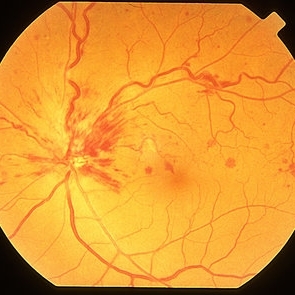 Non Ischemic Hemi-CRVO
Mar 29 2013 by Henry J. Kaplan, MD
Non-ischemic CRVO: blurred disc margins, dilated and tortous veins and scattered hemorrhages in the superior half of the retina.
Condition/keywords: branch retinal vein occlusion (BRVO), central retinal vein occlusion (CRVO), hemi CRVO, non-ischemic central retinal vein occlusion (CRVO)
-
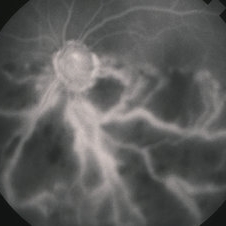
 Inferior Hemi CRVO FFA
Jun 2 2014 by Neha Goel, MS DNB FRCS (Glasg)
FFA of the patient showing NVD and inferior CNP areas.
Photographer: Neha Goel
Imaging device: Zeiss Visucam
Condition/keywords: hemi CRVO, hemicentral retinal vein occlusion
-
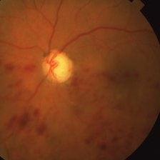
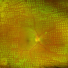
 Superior Hemi-CRVO
Jun 2 2014 by Neha Goel, MS DNB FRCS (Glasg)
Fundus photograph of the right eye of a 55-year-old hypertensive female.
Photographer: Neha Goel
Imaging device: Zeiss Visucam
Condition/keywords: hemi CRVO, hemicentral retinal vein occlusion, retinal hemorrhage
-
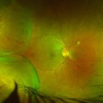
 Superior Hemi CRVO
Aug 31 2014 by Neha Goel, MS DNB FRCS (Glasg)
Fundus photograph of a 60-year-old hypertensive male.
Photographer: Neha Goel
Imaging device: Zeiss Visucam
Condition/keywords: hemi CRVO, hemicentral retinal vein occlusion
-
 Inferior Hemi CRVO
Jun 2 2014 by Neha Goel, MS DNB FRCS (Glasg)
Fundus photograph of the right eye of a 30-year-old hypertensive male.
Photographer: Neha Goel
Imaging device: Zeiss Visucam
Condition/keywords: hemi CRVO, hemicentral retinal vein occlusion
-
 Hemi-CRVO
Mar 27 2019 by Gary R. Cook, MD, FACS
78-year-old African American female patient with COAG and ischemic inferior hemi-CRVO OS; V.A. = HM 1 ft.
Imaging device: Topcon VT-50
Condition/keywords: chronic open-angle glaucoma (COAG), hemi CRVO, retinal hemorrhage
-
 Hemi-CRVO
Jun 4 2019 by Gary R. Cook, MD, FACS
Mid-phase FA image of 78-year-old African American female patient with COAG and a very ischemic inferior hemi-CRVO showing loss of almost all of the capillary bed and staining of the veins in the lower hemisphere of the left eye; V.A. = HM 1 ft.
Imaging device: Topcon VT-50
Condition/keywords: central retinal vein occlusion (CRVO), FA mid phase, fluorescein angiogram (FA), hemi CRVO, ischemia, ischemic CRVO
-
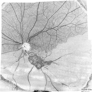
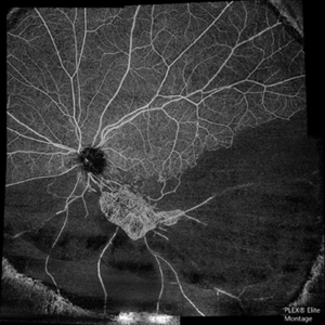 The Barren Field
Jun 26 2020 by SANDEEP KUMAR
A 59-year-old man with DM for 18 years operated for mature cataract. Post op left eye had a visual acuity of 20/80. Wide field swept source OCTA revealed gross vessel wipe out in inferior hemi quadrant with branching out neovascular frond inferior to disc with terminal loops, The patient underwent Anti VEGF injection followed by OCTA guided sectoral retinal photocoagulation.Image J software used here to generate reverse image that sharply delineates the non perfusion are
Photographer: Sandeep Kumar
Imaging device: Optical coherence tomography system Zeiss Plex Elite 9000
Condition/keywords: hemi CRVO
-
 The Barren Field
Jun 27 2020 by SANDEEP KUMAR
A 59-year-old man with DM for 18 years operated for mature cataract. Post op left eye had a visual acuity of 20/80. Wide field swept source OCTA revealed gross vessel wipe out in inferior hemi quadrant with branching out neovascular frond inferior to disc with terminal loops, The patient underwent Anti VEGF injection followed by OCTA guided sectoral retinal photocoagulation.Image J software used here to generate reverse image that sharply delineates the non perfusion area.
Photographer: Sandeep Kumar
Imaging device: Optical coherence tomography system Zeiss Plex Elite 9000
Condition/keywords: hemi CRVO, neovascularization elsewhere (NVE)
-
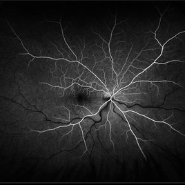
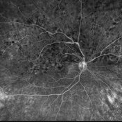 HRVOFA
Aug 13 2021 by Jeffrey Barker
Hemi-retinal vein occlusion. Right-eye.
Photographer: Jeffrey P. Barker, B.S. Retina Vitreous Surgeons of C.N.Y.
Condition/keywords: central retinal vein occlusion (CRVO), hemi CRVO
-
 Fresh lasered retina
Oct 26 2024 by rahul saradge
53 year old male patient was presented with a complaints of diminished vision in LE since 1 month. The BCVA in RE was 6/36p and LE was CF 1/2m. Ocular dilated examination showed RE temporal CD with ?CRVO,OIS and OS showed TRD and old Hemi CRVO. Patient was injected with PST tricot followed by PRP laser at an interval of 1 week. Patient improved to BCVA 6/9.
Photographer: Sakshi Naikade, Isha Netralaya ,Thane
Imaging device: OPTOS
Condition/keywords: crvo, ois, optos, panretinal photo coagulation
-
 Combined Pathology
Oct 26 2024 by rahul saradge
53 year old male patient was presented with a complaints of diminished vision in LE since 1 month. The BCVA in RE was 6/36p and LE was CF 1/2m. Ocular dilated examination showed RE temporal CD with ?CRVO,OIS and OS showed TRD and old Hemi CRVO. Patient was injected with PST tricot followed by PRP laser at an interval of 1 week. Patient improved to BCVA 6/9.
Photographer: Aishwarya Bangar Isha Netralaya Thane
Imaging device: optos
Condition/keywords: choroidal detachment, crvo, ois, optos, pan retinal photocoagulation, tractional retinal detachment
-
 Hemiretinal Vein Occlusion
Nov 14 2024 by Brandon I Fram, MD, BS
40 year-old male with vision changes and observed hemiretinal vein occlusion.
Condition/keywords: branch retinal vein occlusion (BRVO), fluorescein angiogram (FA), Fluorescein angiography, hemi CRVO, hemicentral retinal vein occlusion
 Loading…