Search results (83 results)
-
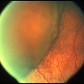
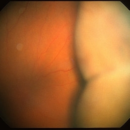
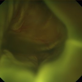
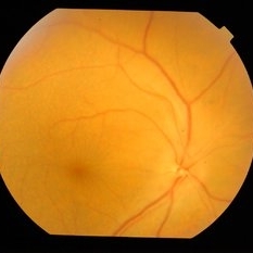
 Choroidal Detachment, In Stereo
Sep 25 2012 by Michael P. Kelly, FOPS
Photographer: Michael P. Kelly, FOPS Director, Duke Eye Labs, Duke University Hospital, Duke Eye Center, Durham, NC
Imaging device: Zeiss FF3C
Condition/keywords: choroidal detachment, stereo pair
-
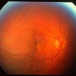
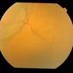
 Choroidal Detachment
Mar 29 2013 by Henry J. Kaplan, MD
One quadrant choroidal detachment as brownish convex lesion.
Condition/keywords: choroidal detachment
-
---thumb.jpg/image-square;max$300,300.ImageHandler) Retina Flower 1
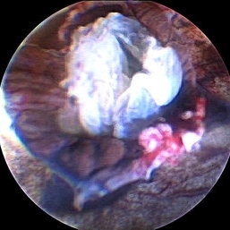
Retina Flower 1
Mar 22 2013 by Cesare Forlini, MD
Post-traumatic Total Choroidal and Retinal Detachment: "like a rose on the rock."
Photographer: Matteo Forlini MD, University of Modena, Institute of Ophthalmology, Modena, Italy
Condition/keywords: choroidal detachment
-
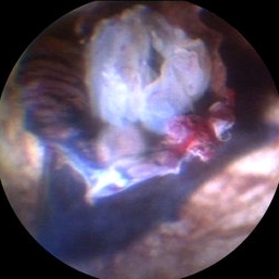
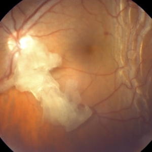 Retained Lens Fragment
Mar 2 2014 by Homayoun Tabandeh, MD, FASRS
Retained lens fragment, choroidal detachment, and serous retinal detachment post cataract surgery
Condition/keywords: retained lens fragments
-
 24 Hours Post Scleral Wound Closure+ Scleral Buckle+25 g Vitrectomy+Silicon Oil
Jan 23 2015 by Carlos Quezada-Ruiz, MD, FASRS
24 hours post op fundus photograph of a 43-year-old man who had perforating injury to the right eye with a small piece of plastic while he was hammering. OD LP, subconjunctival hemorrhage, clear cornea, hyphema, irido and ciclodyalisis as well as a luxated lens with traumatic cataract and a dense vitreous hemorrhage. B-US showed rhegmatogenous retinal detachment with a tear and a big inferior hemorrhagic choroidal detachment. 360 peritomy revealed 2-entry scleral wounds were found in zone II (M V and M VI) and closure was performed. 25 G PPV was performed with the infusion canal placed in the AC through the limbus. Lensectomy and removal of a dense recent vitreous hemorrhage revealed a white detached retina with an exit wound through the temporal inferior segment of the optic nerve with a nasal GRT and sub retinal hemorrhage as well as temporal inferior choroidal, PVD was induced and PFOs helped stabilizing the retina while vitrectomy and sub-retinal hemorrhage was removed through the GRT. Fluid air exchange was made and 360 endolaser over the buckle indentation was done and silicon oil was used as endotamponade. This picture was taken 24 hrs after the surgery.
Photographer: Lilibeth Rodriguez, Instituto de la Visión. Torreon, Mexico.
Condition/keywords: central retinal artery occlusion (CRAO), giant retinal tear, trauma
-
 Choroidal Detachment
Oct 11 2012 by Jeffrey G. Gross, MD, FASRS
Choroidal detachment.
Condition/keywords: choroidal detachment
-
 Choroidal Detachment
Oct 4 2018 by Emily Cooper
Optos photograph of an 80-year-old man presenting with red, painful eye after heart surgery.
Photographer: Emily Cooper, Retina Specialists of Michigan, Grand Rapids MI
Imaging device: Optos
Condition/keywords: choroidal detachment, posterior scleritis
-
 Ocular Hypotony Due to Leaking Bleb
Apr 1 2019 by Anfisa Ayalon, MD
81-year-old male who had trabeculectomy in his right eye 4 years ago, presented to the emergency room with complains of decreased vision in that eye for two months. Slit-lamp examination showed cystic bleb with leakage, intraocular pressure was 0 MMHg. Fundus examination showed hypotony maculopathy, peripheral choroidal detachments, multiple chorioretinal folds with subretinal fluid.
Photographer: Anfisa Ayalon, MD., Meir Medical Center, Kfar Saba, Israel.
Imaging device: California, Optos 200 DTX
Condition/keywords: choroidal detachment, hypotonous retinopathy, hypotony maculopathy
-
 Choroidal Detachment
Mar 29 2013 by Henry J. Kaplan, MD
Choroidal detachment as two adjacent brownish convex lesions.
Condition/keywords: choroidal detachment
-
 Choroidal Detachment
Mar 29 2013 by Henry J. Kaplan, MD
Mild choroidal detachment temporally, macula invived.
Condition/keywords: choroidal detachment
-
 Retina Flower 3
Mar 22 2013 by Cesare Forlini, MD
Post-traumatic Total Choroidal and Retinal Detachment: "like a rose on the rock."
Photographer: Matteo Forlini MD, University of Modena, Institute of Ophthalmology, Modena, Italy
Condition/keywords: choroidal detachment
-
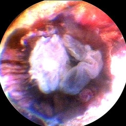 Retina Flower 2
Mar 22 2013 by Cesare Forlini, MD
Post-traumatic Total Choroidal and Retinal Detachment: "like a rose on the rock."
Photographer: Matteo Forlini MD, University of Modena, Institute of Ophthalmology, Modena, Italy
Condition/keywords: choroidal detachment
-
 Choroidal Detachment
Sep 10 2014 by Mehul A Shah
A male patient 5-years-old presented to outdoor and found to have retinal detachment with choroidal detachment following blunt trauma
Photographer: Drashti Netralaya,Dahod
Imaging device: FF 450
Condition/keywords: choroidal detachment
-
 Retina Flower 4
Mar 22 2013 by Cesare Forlini, MD
Post-traumatic Total Choroidal and Retinal Detachment: "like a rose on the rock."
Photographer: Matteo Forlini MD, University of Modena, Institute of Ophthalmology, Modena, Italy
Condition/keywords: choroidal detachment
-
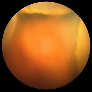
 Suprachoroidal Hemorrhage
Sep 2 2020 by Rinal Pandit
Fundus photograph of left eye of a 56-year-old female with primary angle closure glaucoma showing massive hemorrhagic choroidal detachment that developed following trabeculectomy surgery. Suprachoroidal hemorrhage is defined as the accumulation of blood within the potential space between the choroid and sclera, with the source of the blood being the long or short posterior ciliary artery. Delayed suprachoroidal hemorrhage (DSHC) remains one of the most dreaded and sight threatening complications of glaucoma filtration surgery. The risk factors include old age, hypertension, high myopia, arteriosclerosis, chronically elevated IOP, sudden hypotony, trauma, aphakia/pseudophakia, prior vitrectomy, history of 5 FU injections and anti-platelet agents. The incidence of postoperative SCH after trabeculectomy varies between 0.6%- 1.4%. DSCH after surgery varies considerably in severity but is generally characterized by the sudden onset of severe pain, decreased vision, and a shallow anterior chamber usually associated with raised intraocular pressure. B-scan ultrasonography can help to distinguish serous from hemorrhagic choroidals.Suprachoroidal hemorrhages appear as dome-shaped elevations of the retina with increased echo densities that are often heterogeneous and within the suprachoroidal space. Choroidal effusions appear as dome-shaped elevations with hypoechoic suprachoroidal space. The first step in the management is the timely diagnosis. Medical management includes oral and topical antiglaucoma drugs to lower IOP, oral and topical steroids to control inflammation and topical cycloplegics and oral analgesics to tackle pain. Serial ultrasound B scans of the affected eye should be performed in order to monitor progression of the SCH and help determine apposition, height, and liquefaction of the SCH. Indications of surgical drainage include non resolution with medical management,concurrent retinal detachment, central retinal apposition (kissing choroidals) and incarceration of vitreous in the wound site. The ideal time of drainage is between 7-14 days depending upon clot lysis. The prognosis of both intraoperative and postoperative SCH is poor. An overwhelming majority of patients do not achieve pre-hemorrhage visual acuity and most do not recover to a visual acuity of 20/200 or better. The major determinants of good or bad visual outcomes of SCH’s are preoperative visual acuity and retinal detachment at the time of hemorrhage, respectively.
Imaging device: OPTOS,Ultra wide field retinal imaging system
Condition/keywords: suprachoroidal hemorrhage, trabeculectomy, ultra-wide field imaging
-
 Superchoroid Hemorrhage
Feb 12 2015 by H. Michael Lambert, MD
Choroidal detachment.
Condition/keywords: choroidal detachment, choroidal hemorrhage
-
 Hypotony Maculopathy
Apr 1 2019 by Anfisa Ayalon, MD
Fundus autofluorescence image of 81-year-old male with right eye ocular hypotony due to leaking bleb. Note severe hypotony maculopathy, peripheral choroidal detachments, multiple chorioretinal folds.
Photographer: Anfisa Ayalon, MD., Meir Medical Center, Kfar Saba, Israel.
Imaging device: California, Optos 200 DTX
Condition/keywords: choroidal detachment, choroidal folds, fundus autofluorescence (FAF), hypotonous retinopathy, hypotony maculopathy
-
 Superchoroid Hemorrhage
Feb 12 2015 by H. Michael Lambert, MD
A-scan shows choroidal detachment with hemorrhage.
Condition/keywords: choroidal detachment, choroidal hemorrhage
-
 Superchoroid Hemorrhage
Feb 12 2015 by H. Michael Lambert, MD
Choroid detachment with vitreous hemorrhage.
Condition/keywords: choroidal detachment, choroidal hemorrhage
-
 Superchoroid Hemorrhage
Feb 12 2015 by H. Michael Lambert, MD
B-scan shows choroidal detachment with hemorrhage.
Condition/keywords: choroidal detachment, hemorrhage
-
 Uveitis With Exudative Retinal Detachment
May 3 2014 by Mallika Goyal, MD
Fundus photograph of an elderly patient with bilateral posterior uveitis and subnormal vision. Posterior pole looks relatively unremarkable though periphery reveal s choroidal detachments and exudative retinal detachment. Fluorescein angiogram reveals punctate hyperfluorecence through the entire fundus and disc hyperfluorescence.
Photographer: Mallika Goyal, MD, Apollo Health City, Jubilee Hills, Hyderabad, India
Condition/keywords: exudative retinal detachment, uveitis
-
 Choroidal Detachments
Aug 11 2015 by H. Michael Lambert, MD
Ultrasound of choroidal detachments.
Condition/keywords: choroidal detachment, ultrasound
-
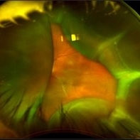
 Kissing Serous Choroidal Detachment
Mar 8 2023 by Annaka Gooding
Ultra-widefield fundus photograph of a 73 year old male with a Kissing serous choroidal detachment affecting his right eye. Patient presented at the office following a XEN implant and his vision was sc20/100 PH20/50+1. The physician recommended to start Prednisone treatment.
Photographer: Annaka Gooding
Imaging device: Optos California
Condition/keywords: fundus photography, Kissing Serous Choroidal Detachment, Optos, Right Eye, ultra-wide field imaging
-
 Uveitis With Exudative Retinal Detachment
May 3 2014 by Mallika Goyal, MD
Choroidal detachment in an elderly male patient with bilateral uveitis. B-scan did not reveal underlying tumor. MRI brain to rule out lymphoma was unremarkable. Response to oral steroids was good with complete resolution of the choroidal detachment and other features.
Photographer: Mallika Goyal, MD, Apollo Health City, Jubilee Hills, Hyderabad, India
Condition/keywords: exudative retinal detachment, uveitis
-
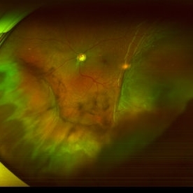
 Traumatic Giant Retinal Tear Associated Retinal Detachment
Nov 9 2019 by Luis J Haddock, MD
This wide field fundus photograph of the left eye shows a traumatic giant retinal tear associated with total retinal detachment. The image shows the torn superior retina folded over the macula with the underside of the retina visible. There is associated peripheral choroidal detachment due to hypotony from giant retinal tear. This patient has history of spondyloepithelial dysplasia with dwarfism and presented with vision loss after a recent blunt trauma with elbow to the eye.
Imaging device: Optos
Condition/keywords: giant retinal tear, traumatic optic neuropathy
 Loading…