Search results (17 results)
-
---thumb.jpg/image-square;max$300,300.ImageHandler) Nummular Depigmented Or Atrophic Pigment Epithelium
Nummular Depigmented Or Atrophic Pigment Epithelium
Oct 24 2013 by Maurice F. Rabb
61 year old with nummular areas peripherally of depigmented or atrophic pigment epithelium with pigmented bridges between the nummular (circular) areas, completely encircling the posterior equator from the level of the posterior arcades out to the equator.
Condition/keywords: atrophic pigment epithelium, nummular depigmented epithelium
-
 Tilted Disc Syndrome Complicated with RPE Atrophy and Polypoidal Choroidal Vasculopathy
Jan 20 2020 by Pierre-Henry Gabrielle, MD
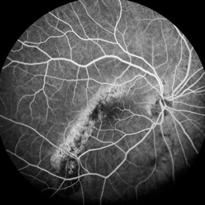
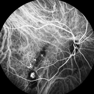
Early-phase FA angiography of an 81-year-old woman with a tilted disc syndrome complicated with RPE atrophy and polypoidal choroidal vasculopathy.
Photographer: Pierre-Henry Gabrielle, Ophthalmology department, Dijon University Hospital, France.
Imaging device: Heidelberg spectralis angiography
Condition/keywords: atrophic pigment epithelium, fluorescein angiogram (FA), polypoidal choroidal vasculopathy (PCV), tilted disc
-
 Atrophic Pigment Epithelium
Jul 19 2019 by JEFFERSON R SOUSA, Tecg.º (Biomedical Systems Technology)
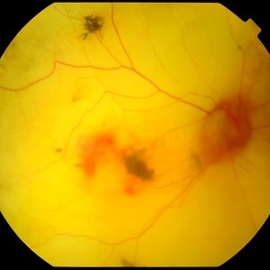
Patient 16-years-old, visual acuity with light perception. In retinal evaluation presented total atrophy retinal pigment epithelium and total papilla excavation. Mobilization of pigments and presence of macular hemorrhage.
Photographer: JEFFERSON R SOUSA - Study Center and Ophthalmological Research Dr. Andre M V Gomes, Institute Dr. Suel Abujamra São Paulo-Brazil
Imaging device: Topcon TRC-50 DX, Imaginet 4.0, angle de 35 graus. Flash 12w-s
Condition/keywords: atrophic pigment epithelium
-
 Tilted Disc Syndrome Complicated with RPE Atrophy and Polypoidal Choroidal Vasculopathy
Jan 20 2020 by Pierre-Henry Gabrielle, MD
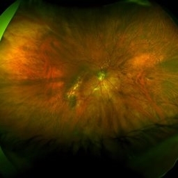
Fundus optos photograph of an 81-year-old woman with a tilted disc syndrome complicated with RPE atrophy and polypoidal choroidal vasculopathy.
Photographer: Pierre-Henry Gabrielle, Ophthalmology department, Dijon University Hospital, France.
Imaging device: Optos
Condition/keywords: atrophic pigment epithelium, indocyanine green (ICG) angiography, polypoidal choroidal vasculopathy (PCV), tilted disc
-
 Tilted Disc Syndrome Complicated with RPE Atrophy and Polypoidal Choroidal Vasculopathy
Jan 20 2020 by Pierre-Henry Gabrielle, MD
Zoomed fundus optos photograph of an 81-year-old woman with a tilted disc syndrome complicated with RPE atrophy and polypoidal choroidal vasculopathy.
Photographer: Pierre-Henry Gabrielle, Ophthalmology department, Dijon University Hospital, France.
Imaging device: Optos
Condition/keywords: atrophic pigment epithelium, Optos, polypoidal choroidal vasculopathy (PCV), tilted disc
-
 Tilted Disc Syndrome Complicated with RPE Atrophy and Polypoidal Choroidal Vasculopathy
Jan 20 2020 by Pierre-Henry Gabrielle, MD
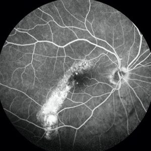
Late-phase fluorescein angiography of an 81-year-old woman with a tilted disc syndrome complicated with RPE atrophy and polypoidal choroidal vasculopathy.
Photographer: Pierre-Henry Gabrielle, Ophthalmology department, Dijon University Hospital, France.
Imaging device: Heidelberg spectralis angiography
Condition/keywords: atrophic pigment epithelium, fluorescein angiogram (FA), polypoidal choroidal vasculopathy (PCV), tilted disc
-
 Tilted Disc Syndrome Complicated with RPE Atrophy and Polypoidal Choroidal Vasculopathy
Jan 20 2020 by Pierre-Henry Gabrielle, MD
Coupled OCT B-scan and ICG angiography of an 81-year-old woman with a tilted disc syndrome complicated with RPE atrophy and polypoidal choroidal vasculopathy.
Photographer: Pierre-Henry Gabrielle, Ophthalmology department, Dijon University Hospital, France.
Imaging device: Heidelberg spectralis
Condition/keywords: atrophic pigment epithelium, indocyanine green (ICG) angiography, optical coherence tomography (OCT), polypoidal choroidal vasculopathy (PCV), tilted disc
-
 Tilted Disc Syndrome Complicated with RPE Atrophy and Polypoidal Choroidal Vasculopathy
Jan 20 2020 by Pierre-Henry Gabrielle, MD
Early-phase ICG angiography of an 81-year-old woman with a tilted disc syndrome complicated with RPE atrophy and polypoidal choroidal vasculopathy.
Photographer: Pierre-Henry Gabrielle, Ophthalmology department, Dijon University Hospital, France.
Imaging device: Heidelberg spectralis angiography
Condition/keywords: atrophic pigment epithelium, indocyanine green (ICG) angiography, polypoidal choroidal vasculopathy (PCV), tilted disc
-
 Tilted Disc Syndrome Complicated with RPE Atrophy and Polypoidal Choroidal Vasculopathy
Jan 20 2020 by Pierre-Henry Gabrielle, MD
Coupled OCT B-scan and ICG angiography of an 81-year-old woman with a tilted disc syndrome complicated with RPE atrophy and polypoidal choroidal vasculopathy.
Photographer: Pierre-Henry Gabrielle, Ophthalmology department, Dijon University Hospital, France.
Imaging device: Heidelberg spectralis
Condition/keywords: atrophic pigment epithelium, indocyanine green (ICG) angiography, optical coherence tomography (OCT), polypoidal choroidal vasculopathy (PCV), tilted disc
-
 Tilted Disc Syndrome Complicated with RPE Atrophy and Polypoidal Choroidal Vasculopathy
Jan 20 2020 by Pierre-Henry Gabrielle, MD
Late-phase ICG angiography of an 81-year-old woman with a tilted disc syndrome complicated with RPE atrophy and polypoidal choroidal vasculopathy.
Photographer: Pierre-Henry Gabrielle, Ophthalmology department, Dijon University Hospital, France.
Imaging device: Heidelberg spectralis angiography
Condition/keywords: atrophic pigment epithelium, indocyanine green (ICG) angiography, polypoidal choroidal vasculopathy (PCV), tilted disc
-
 Tilted Disc Syndrome Complicated with RPE Atrophy and Polypoidal Choroidal Vasculopathy
Jan 20 2020 by Pierre-Henry Gabrielle, MD
Coupled OCT B-scan and ICG angiography of an 81-year-old woman with a tilted disc syndrome complicated with RPE atrophy and polypoidal choroidal vasculopathy.
Photographer: Pierre-Henry Gabrielle, Ophthalmology department, Dijon University Hospital, France.
Imaging device: Heidelberg spectralis
Condition/keywords: atrophic pigment epithelium, indocyanine green (ICG) angiography, optical coherence tomography (OCT), polypoidal choroidal vasculopathy (PCV), tilted disc
 Loading…