Search results (27 results)
-
 AVM
AVM
-
 Vascular Anormalities
Jan 6 2016 by Andrea Arriola-Lopez, MD MSc
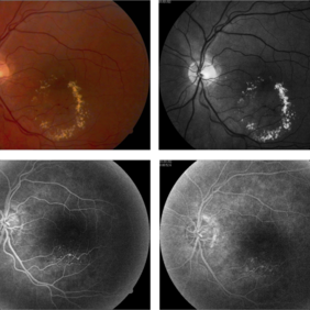
77-year-old man. Decrease of visual acuity OS. VA 20/30 IOP 14mmHg. Fundus examination findings: Hard exudates, microaneurysms near to fovea. OCT shows IRF. Late leakage on FA.
Photographer: Andrea Elizabeth Arriola-Lopez, MSc MD
Condition/keywords: abnormal retinal vessel, aneurysm, hard exudates, vascular anomaly
-
 Coats' Disease - Stage 3A
Aug 21 2019 by Victor M Villegas, MD
Coats' Disease - stage 3A.
Condition/keywords: abnormal retina, Coats' disease, diffuse lipid exudate, edema, foveal hard exudates, pediatic retina, retcam, retinal angioma
-
 AVM
AVM
-
 AVM
AVM
Aug 21 2013 by Howard Schatz, MD
Twenty year old white female, 20/25 OU.
Condition/keywords: abnormal retinal vessel
-
 AVM
AVM
-
 AVM
AVM
Aug 21 2013 by Howard Schatz, MD
Fifty eight year old white female.
Condition/keywords: abnormal retinal vessel
-
 AVM
AVM
Aug 21 2013 by Howard Schatz, MD
Not an AV shunt, Idiopathic RV, superior nasal grads.
Condition/keywords: abnormal retinal vessel
-
 AVM
AVM
Aug 21 2013 by Howard Schatz, MD
Sixty year old white female, right eye 20/20, left eye 10/200, venous loop.
Condition/keywords: abnormal retinal vessel
-
 AVM
AVM
-
 AVM
AVM
Aug 21 2013 by Howard Schatz, MD
Twenty three year old white male, 20/20 OU.
Condition/keywords: abnormal retinal vessel
-
 AVM
AVM
-
 AVM
AVM
Aug 21 2013 by Howard Schatz, MD
Twenty seven year old white male, right eye 20/20, left eye 20/20.
Condition/keywords: abnormal retinal vessel
-
 AVM
AVM
Aug 21 2013 by Howard Schatz, MD
Twenty five year old white female, right eye 20/20, left eye 20/80.
Condition/keywords: abnormal retinal vessel
-
 AVM
AVM
Aug 21 2013 by Howard Schatz, MD
Thirty eight year old female, right eye 20/20, left eye 20/200.
Condition/keywords: abnormal retinal vessel
-
 IJFT
IJFT
Mar 26 2018 by Purva Patwari
Defective vision.
Photographer: Dr Purva Patwari, Patwari Retina Center, Ahmedabad, Gujarat , India
Condition/keywords: abnormal retinal vessel, IJFT, IJT
-
 Advanced Coats' Disease with Neovascular Glaucoma
Aug 21 2019 by Victor M Villegas, MD
Advanced Coats' Disease with neovascular glaucoma.
Photographer: Giselle Deoliveira, Bascom Palmer Eye Institute, University of Miami
Imaging device: RetCam III
Condition/keywords: abnormal retinal vessel, bullous retinal detachment, Coats' disease, diffuse lipid exudate, foveal hard exudates, neovascular glaucoma, pediatric retina
-
 Sunset Glow Fundus
May 15 2022 by Manuel Ángel Alcántara Delgado, MD
Optomap ultra-widefield retinal imaging of an 35-year-old woman showed sunset glow fundus, multiple nummular chorioretinal atrophic lesions, macular subretinal fibrosis and pigment clumping in chronic recurrent stage of Vogt-Koyanagi-Harada disease.
Photographer: Manuel Ángel Alcántara Delgado. Conde de Valenciana.
Condition/keywords: abnormal retina, benign pigmented lesions, pigment clumps, retinal fibrosis, uveitis, Vogt-Koyanagi-Harada
-
 AVM
AVM
-
 Spontaneously dropped Nucleus In a Congenital Rubella Retina disease
Apr 29 2022 by NEIFFER RABELO
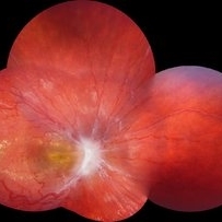
Intraoperative Fundus Photograph of an 68 years old with past medical history of
Photographer: Rodrigo Dos Anjos Vesiani, Retina Institute - Belo Horizonte - Brazil
Imaging device: OPMI LUMERA® 700
Condition/keywords: abnormal retina, dropped nucleus, retina surgery, rubella
-
 Combined Hamartoma of Retina and RPE
Jul 15 2020 by Itzel Ocampo
Fundus photograph of a 53-year-old man with a combined hamartoma of retina and RPE; visual capacity of hand movement. No systemic associations.
Photographer: Itzel Ocampo, Universidad Autonoma de Mexico, Hospital General de Mexico "Eduardo Liceaga"
Condition/keywords: abnormal retina, combined hamartoma
-
 Slide 13-28
Mar 4 2019 by Lancaster Course in Ophthalmology
Pseudoglioma: abnormal retina behind the lens with tunica vasculosa lentis in a maldeveloped eye associated with chromosomal defect ( x25).
Condition/keywords: abnormal retina, chromosomal defect, pseudoglioma, tunica vasculosa lentis
-
 Retinal Capillary Hemangioma
Sep 9 2021 by Jesus Lozano, MD
60 year-old woman with a Peripheral RCH treated with laser photocoagulation.
Photographer: Yair Bet Yosef, Hadassah Medical Center. Israel
Imaging device: Optos Silverstone
Condition/keywords: abnormal retinal vessel, anomalous vessels, dilated tortuous vessels, hemangioma, retina
-
 Von Hippel Lindau with retinal capillary hemangioma
Nov 2 2023 by Marcelo Zas, MD PhD
30-year-old female patient diagnosed with Syndrome VHL (Von Hippel Lindau). Stage II. In the first wide-field retinography of the right eye we can observe the exophytic retinal hemangiomas, rounded, slightly delimited, located in the peripheral retina in the upper and lower temporal quadrants and due to the exudation produced by them, hard exudates are observed in the star hemisphere, affecting the macula.
Photographer: Mariano Cotic MD
Imaging device: Silverstone SS OCT Optos
Condition/keywords: abnormal retinal vessel
-
 Presumed Combined Hamartoma of the Retina and the Retinal Pigment Epithelium
Dec 7 2020 by Martin J Siemerink, MD, PhD
7-year-old girl, right eye VA 20/200, left eye unremarkable.
Photographer: Faroch Payman, Bergman Clinics Doetinchem
Condition/keywords: abnormal retinal vessel, combined hamartoma
 Loading…