Search results (11 results)
-
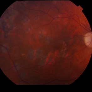
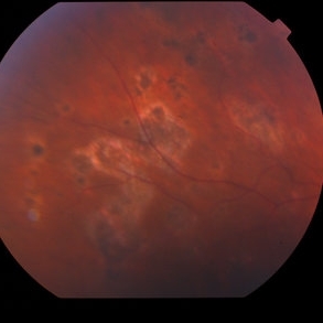 West Nile Virus Choroiditis
Apr 4 2014 by Suber S. Huang, MD, MBA, FASRS
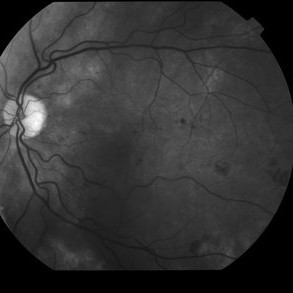
Fundus photograph (11 month follow-up) of an 88-year-old woman who developed West Nile virus encephalitis on 08/2012 and subsequent choroiditis.
Photographer: Geoffrey Pankhurst; University Hospitals Eye Institute, Case Western Reserve University, Cleveland, OH
Imaging device: TopCon TRC50EX
Condition/keywords: choroiditis, disseminated choroiditis, infectious uveitis, optic nerve atrophy
-
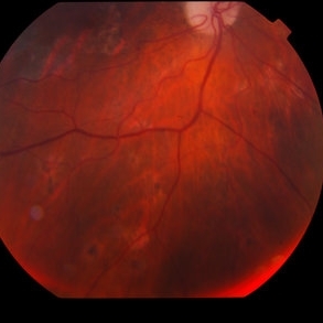
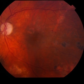 West Nile Virus Choroiditis
Apr 4 2014 by Suber S. Huang, MD, MBA, FASRS
Fundus photograph (11 month follow-up) of an 88-year-old woman who developed West Nile virus encephalitis on 08/2012 and subsequent choroiditis.
Photographer: Geoffrey Pankhurst; University Hospitals Eye Institute, Case Western Reserve University, Cleveland, OH
Imaging device: TopCon TRC50EX
Condition/keywords: choroiditis, disseminated choroiditis, infectious uveitis, optic nerve atrophy
-
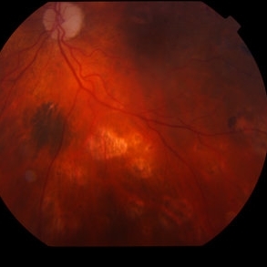
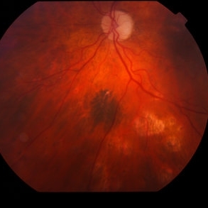 West Nile Virus Choroiditis
Apr 4 2014 by Suber S. Huang, MD, MBA, FASRS
Fundus photograph (11 month follow-up) of an 88-year-old woman who developed West Nile virus encephalitis on 08/2012 and subsequent choroiditis.
Photographer: Geoffrey Pankhurst; University Hospitals Eye Institute, Case Western Reserve University, Cleveland, OH
Imaging device: TopCon TRC50EX
Condition/keywords: choroiditis, disseminated choroiditis, infectious uveitis, optic nerve atrophy
-
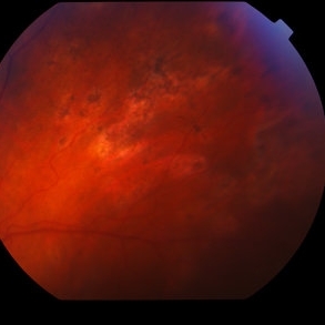
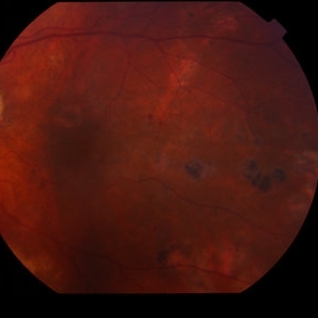 West Nile Virus Choroiditis
Apr 4 2014 by Suber S. Huang, MD, MBA, FASRS
Fundus photograph (11 month follow-up) of an 88-year-old woman who developed West Nile virus encephalitis on 08/2012 and subsequent choroiditis.
Photographer: Geoffrey Pankhurst; University Hospitals Eye Institute, Case Western Reserve University, Cleveland, OH
Imaging device: TopCon TRC50EX
Condition/keywords: choroiditis, disseminated choroiditis, infectious uveitis, optic nerve atrophy
-
 West Nile virus choroiditis
Apr 4 2014 by Suber S. Huang, MD, MBA, FASRS
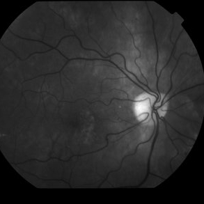
Fundus photograph (11 month follow-up) of an 88-year-old woman who developed West Nile virus encephalitis on 08/2012 and subsequent choroiditis
Photographer: Geoffrey Pankhurst; University Hospitals Eye Institute, Case Western Reserve University, Cleveland, OH
Imaging device: TopCon TRC50EX
Condition/keywords: choroiditis, disseminated choroiditis, infectious uveitis, optic nerve atrophy, West Nile virus choroiditis
-
 West Nile Virus Choroiditis
Apr 4 2014 by Suber S. Huang, MD, MBA, FASRS
Fundus photograph (11 month follow-up) of an 88-year-old woman who developed West Nile virus encephalitis on 08/2012 and subsequent choroiditis.
Photographer: Geoffrey Pankhurst; University Hospitals Eye Institute, Case Western Reserve University, Cleveland, OH
Imaging device: TopCon TRC50EX
Condition/keywords: choroiditis, disseminated choroiditis, infectious uveitis, optic nerve atrophy
-
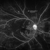 West Nile Virus Retinopathy with Retinal Vascular Occlusion
Nov 19 2019 by Ravi Keshavamurthy, MBBS,MD
39-year-old male with history of recent hospitalization for West Nile Virus encephalitis had vision loss in right eye. Examination revealed characteristic curvilinear chorioretinitis patches on fluorescein angiogram with retinal artery occlusion.
Imaging device: Optos
Condition/keywords: West Nile virus choroiditis
 Loading…