Search results (65 results)
-
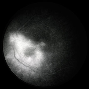
 Cystoid Macular Edema
Oct 8 2012 by Jeffrey G. Gross, MD, FASRS
CME, s/p AC-IOL, FA late phase.
Condition/keywords: cystoid macular edema (CME), late phase
-
 Sickle SC Sea Fan
Oct 8 2012 by Jeffrey G. Gross, MD, FASRS
Sickle SC sea fan, partial regression, FA late phase leakage.
Condition/keywords: FA late phase leakage, partial regression, sea fan, sickle cell
-
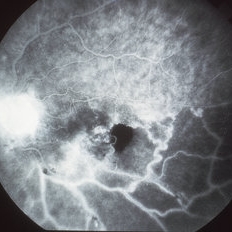
 Toxoplasmosis Chorioretinitis
Oct 10 2012 by Jeffrey G. Gross, MD, FASRS
Toxoplasmosis chorioretinitis, 20/400, + APD, FA late phase.
Condition/keywords: 20/400, afferent pupillary defect (APD), FA late phase, toxoplasmosis chorioretinitis
-
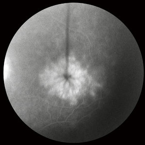
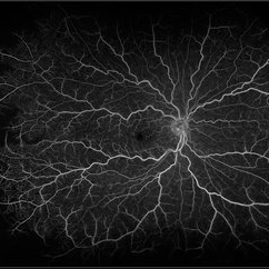
 FA Late Phase Optic Disc Edema
Oct 1 2012 by Jeffrey G. Gross, MD, FASRS
FA late phase optic disc edema with disc leakage in patient with subdural hematoma.
Condition/keywords: disc leakage, subdural hematoma
-
 CME
CME
May 3 2013 by Suber S. Huang, MD, MBA, FASRS
CME.
Imaging device: Retina Diseases Imaging Analysis Reading Center
Condition/keywords: angiographic macular leakage, cystoid macular edema (CME), FA late phase leakage
-
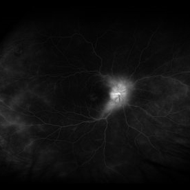
 CRVO with Choroidal Profusion Delay
Oct 18 2012 by Raj K. Maturi, MD
Photographer: Tom Steele, CRA
Imaging device: Topcon 50dx
Condition/keywords: choroidal profusion delay, FA late phase
-
 Behcet's Eye Disease
Apr 10 2017 by Deepak Bhojwani, MS
A 19-year-old boy presented with with recurrent oral and genital ulcers along with blurring of vision. Systemically he was HLA B-51 Positive suggesting Behcet's Disease. The FFA photograph depicts the classic small vessel immune mediated vasculitis predominantly affecting the capillaries (capillaropathy).
Photographer: DEEPAK BHOJWANI, RAGHUDEEP EYE HOSPITAL, AHMEDABAD.
Condition/keywords: Behcet's Disease, Behcet's uveitis, FA late phase leakage
-
 Macular Telangiectasia (FA Late Phase)
May 16 2014 by Avris Romario Diparaja Siahaan
FA (late phase) image of a 58-year-old-man with a macular telangiectasia condition on his left eye.
Photographer: Avris Romario Diparaja Siahaan, Klinik Mata Nusantara
Imaging device: Topcon TRC 50 DX Type IA
Condition/keywords: FA late phase, macular telangiectasia
-
 ROP FA OS
Apr 27 2018 by Brenda Fallas
4-month-old baby with regressed ROP post-Avastin.
Photographer: Brenda Fallas, Bascom Palmer Eye Institute, Miami, FL
Imaging device: RETCAM III 130 degree lens montage
Condition/keywords: FA late phase leakage, fluorescein angiogram (FA), retinopathy of prematurity (ROP)
-
 Syphilis Neuroretinopathy FA
Sep 25 2013 by Alexandre Durao Alves Pereira, MD
Syphilis neuroretinopathy, late phase FA.
Photographer: Alexandre Pereira
Condition/keywords: FA late phase
-
---thumb.jpg/image-square;max$300,300.ImageHandler) Pattern Dystrophy
Pattern Dystrophy
Aug 9 2013 by From the Collections of Thomas M. Aaberg, MD and Thomas M. Aaberg Jr., MD
Late FA of patient with pattern dystrophy.
Condition/keywords: FA late phase, pattern macular dystrophy
-
 Polypoidal Choroidal Vasculopathy (FA Late Phase)
May 16 2014 by Avris Romario Diparaja Siahaan
FA image (late phase) a 66-year-old woman with a polypoidal choroidal vasculopathy (PCV) on her right eye. She had a injection (Avastin) for several times.
Photographer: Avris Romario Diparaja Siahaan, Klinik Mata Nusantara
Imaging device: Heidelberg HRA + OCT Spectralis
Condition/keywords: FA late phase, polypoidal choroidal vasculopathy (PCV)
-
 ROP FA OD
Apr 27 2018 by Brenda Fallas
4-month-old baby with regressed ROP post-Avastin.
Photographer: Brenda Fallas, Bascom Palmer Eye Institute, Miami, FL
Imaging device: RETCAM III 130 degree lens mongtage
Condition/keywords: FA late phase leakage, fluorescein angiogram (FA), retina, retinopathy of prematurity (ROP)
-
---thumb.jpg/image-square;max$300,300.ImageHandler) Pattern Dystrophy
Pattern Dystrophy
Aug 9 2013 by From the Collections of Thomas M. Aaberg, MD and Thomas M. Aaberg Jr., MD
Late FA of patient with pattern dystrophy.
Condition/keywords: FA late phase, pattern macular dystrophy
-
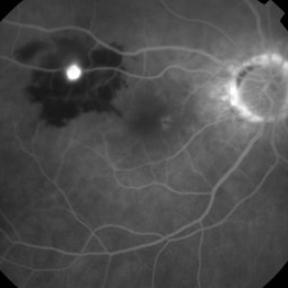
 Von Hippel-Lindau (FA Late Phase)
May 17 2014 by Avris Romario Diparaja Siahaan
Fluorescein angiogram (Late Phase) photograph of a 57-year-old woman with a Von Hippel-Lindau
Photographer: Avris Romario Diparaja Siahaan, Klinik Mata Nusantara
Imaging device: Topcon TRC 50 DX Type IA
Condition/keywords: FA late phase, Von Hippel-Lindau
-
---thumb.jpg/image-square;max$300,300.ImageHandler) Pattern Dystrophy
Pattern Dystrophy
Aug 9 2013 by From the Collections of Thomas M. Aaberg, MD and Thomas M. Aaberg Jr., MD
Left eye, late FA patient with pattern dystrophy.
Condition/keywords: FA late phase, left eye, pattern macular dystrophy
-
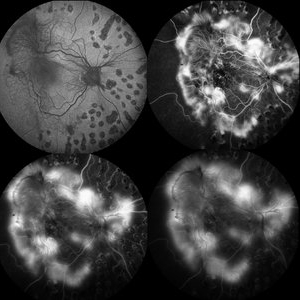 Proliferative Diabetic Retinopathy
May 28 2016 by Olivia Rainey
Fluorescein angiogram series of an 30-year-old male with proliferative diabetic retinopathy affecting his right eye. The patient presented with worsening neovascularization and scar tissue contracting in macula in the right eye. He experienced a decline in vision secondary to macula ischemia. Patient was seeing 20/400 and with PH 20/200 in the right eye and HM in the left eye.
Photographer: Olivia Rainey
Imaging device: Heidelberg Spectralis
Condition/keywords: diabetes, FA early phase, FA late phase, FA mid phase, fluorescein leakage, fundus autofluorescence (FAF), neovascularization (NV), proliferative diabetic retinopathy (PDR)
-
---thumb.jpg/image-square;max$300,300.ImageHandler) Pattern Dystrophy
Pattern Dystrophy
Aug 9 2013 by From the Collections of Thomas M. Aaberg, MD and Thomas M. Aaberg Jr., MD
Late FA, left eye of patient with pattern dystrophy.
Condition/keywords: FA late phase, left eye, pattern macular dystrophy
-
---thumb.jpg/image-square;max$300,300.ImageHandler) Pattern Dystrophy
Pattern Dystrophy
Aug 9 2013 by From the Collections of Thomas M. Aaberg, MD and Thomas M. Aaberg Jr., MD
Right eye, late FA patient with pattern dystrophy.
Condition/keywords: FA late phase, pattern macular dystrophy
-
---thumb.jpg/image-square;max$300,300.ImageHandler) Pattern Dystrophy
Pattern Dystrophy
Aug 9 2013 by From the Collections of Thomas M. Aaberg, MD and Thomas M. Aaberg Jr., MD
Late FA of patient with pattern dystrophy.
Condition/keywords: FA late phase, pattern macular dystrophy
-
 RAMA
RAMA
-
 Central Retinal Vein Occlusion With Macular Edema
Aug 29 2018 by Olivia Rainey
Ultra-widefield fluorescein angiogram of an 58-year-old male with a central retinal vein occlusion with macular edema affecting his right eye. Fluorescein showed delayed transit with late leakage.
Photographer: Olivia Rainey
Imaging device: Optos
Condition/keywords: central retinal vein occlusion (CRVO), FA late phase leakage, fluorescein angiogram (FA), Optos, tortuous vessels, ultra-wide field imaging
-
 Polypoidal Choroidal Vasculopathy
Jul 20 2023 by Gregg T. Kokame, MD, MMM, FASRS
64 Year Old Male, with Polypoidal Choroidal Vasculopathy. Pre-op and Post-op PDT/Vabysmo Injection
Photographer: Jaclyn Pisano
Imaging device: Heidelberg Spectralis
Condition/keywords: FA late phase, indocyanine green (ICG) angiography, OCT, PDT, polypoidal choroidal vasculopathy (PCV), subretinal, subretinal fluid
-
 Syphilitic Uveitis
Apr 2 2020 by Olivia Rainey
Ultrawide-field fluorescein angiogram of a 42-year-old male with syphilitic uveitis affecting his right eye more than his left. Patient is HIV positive. He developed hearing loss and palm/leg/scalp rash prompting diagnosis of neurosyphilis, s/p IM and full IV course of 2.4 Mil PCN G, and finished this course 3/9/20. He admits to recent rectal bleeding with ongoing plan for colonoscopy 3/16/20. He has a history of extensive travel including London, Hong Kong, and Bangkok. His husband has also been treated with IV PCN G, however per chart review he has multiple sexual partners. Patient's vision was 20/20 in each eye.
Photographer: Olivia Rainey
Imaging device: Optos California
Condition/keywords: disc hyperfluorescence, FA late phase leakage, fluorescein angiogram (FA), fluorescein leakage, HIV, late phase, optic nerve edema, Optos, phelbitis, syphilis neuroretinopathy, ultra-wide field imaging, uveitis
-
 Nonperfused BRVO with Collateral Vessels
Apr 8 2019 by Gary R. Cook, MD, FACS
Late-phase fluorescein angiogram image of the left eye of a 73-year-old African-American female with a nonperfused BRVO showing flow through the collateral vessels, marked loss of the capillary bed, disc leakage from some NVD, and ischemic staining of the retinal veins; V.A. = 20/70-1
Imaging device: Topcon VT-50
Condition/keywords: branch retinal vein occlusion (BRVO), capillary nonperfusion, collaterals, disc leakage, FA late phase, fluorescein angiogram (FA)
 Loading…