Search results (6 results)
-
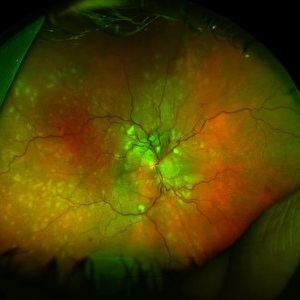
 Hypertensive Choroidopathy - Right Eye
Dec 21 2016 by Maciej Czepita
Fundus photograph and SD-OCT scan as well as fundus autofluorescence image (FAF) of the right eye of a 70-year-old woman with hypertensive choroidopathy. In the fundus image numerous Elschnig's spots are visible. Note the Hollenhorst plaque in the superior temporal artery. In the SD-OCT scan (green line on the fundus image) the RPE layer is uneven. Numerous hypo and hyperautofluorescent patches can be seen in the fundus autofluorescence image.
Photographer: Maciej Czepita, M.D., Ph.D., Pomeranian Medical University, Szczecin, Poland
Imaging device: Heidelberg Spectralis HRA+OCT
Condition/keywords: hypertensive choroidopathy
-
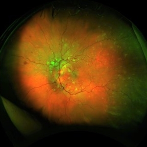
 Hypertensive Retinopathy, Right
Feb 23 2017 by Alla Goldberg, MD
Fundus photograph of 35-year-old man with severe hypertension (182/128).
Photographer: Sofia Rutiaga, UT Health McGovern Medical School, Cizik Eye Clinic
Condition/keywords: cotton wool spots, Elschnig's spots, hypertensive choroidopathy, hypertensive retinopathy, serous retinal detachment
-
 Hypertensive retinopathy, left
Feb 23 2017 by Alla Goldberg, MD
Fundus photograph of 35-year-old man with severe hypertension (182/128).
Photographer: Sofia Rutiaga, UT Health McGovern Medical School, Cizik Eye Clinic
Condition/keywords: cotton wool spots, Elschnig's spots, hypertensive choroidopathy, hypertensive retinopathy, serous retinal detachment
-
 Slide 10-14
Feb 26 2019 by Lancaster Course in Ophthalmology
Regeneration cataract with Elschnig's pearls ( X 16). Similar bladder cells (Wedl cells) are seen beneath the posterior capsule in a posterior subcapsular cataract. These are found after extracapsular extraction. (Scheie Eye Institute, No. 4083.)
Condition/keywords: cataract, Elschnig's spots, Wedl cells
-
 Slide 9-23
Feb 26 2019 by Lancaster Course in Ophthalmology
Choroidal infarct (Elschnig spot). There is a localized area of loss of retinal pigment epithelium and outer ischemic retinal atrophy with loss of the photoreceptor cell and outer plexiform layers, and partial loss of the inner nuclear layer without any reparative changes.
Condition/keywords: choroidal infarction, Elschnig's spots
-
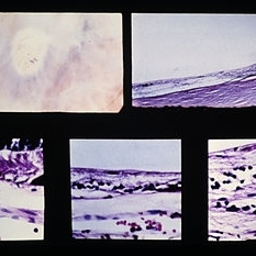 Slide 9-65
Feb 26 2019 by Lancaster Course in Ophthalmology
Elschnig spot. Localized choroidal infarction with loss of choriocapillaris, RPE, and outer layers of the retina. The thinned inner nuclear layer of the retina rests against Bruch's membrane. At the anterior margin (lower left view) there is an abrupt transition (arrow) between the normal area (left) where the choriocapillaris and RPE are intact and the area of post-ischemic atrophy of the structures (right). A similar but reversed configuration is observed at the posterior margin (lower right view).
Condition/keywords: Bruch's membrane, choroidal infarction, Elschnig's spots
 Loading…