Search results (21 results)
-
 Dislocated Lens
Apr 26 2023 by Chloe Hanifan
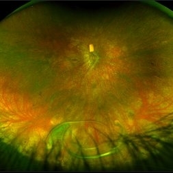
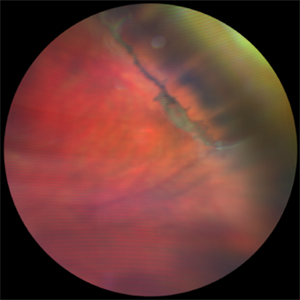
Ultra wide field fundus photograph of a 41-year-old male with a dislocated lens affecting his right eye. IOL noted inferior vitreous base and vitrectomy surgery for removal of IOL was recommended. Patient has history of retinitis pigmentosa as well. Patient's vision at the time of presentation was counting fingers at 2 feet.
Photographer: Chloe Hanifan
Imaging device: Optos California
Condition/keywords: dislocated lens, fundus photography, Optos, pseudocolor, retinitis pigmentosa, ULTRA WIDE FIELD
-
 Vitreous Base Avulsion
Sep 19 2019 by Anfisa Ayalon, MD
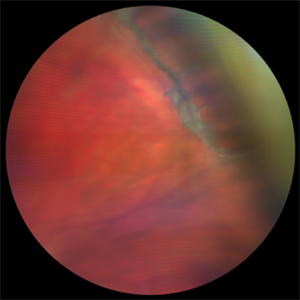
Fundus picture of a 34-year-old patient with left eye vitreous base avulsion three months after rhegmatogenous retinal detachment repair with circular scleral buckle implantation. Note bucket handle sign and 360 degrees scleral buckle indentation with a flat retina.
Photographer: Anfisa Ayalon, MD., Meir Medical Center, Kfar Saba, Israel.
Imaging device: California, Optos 200 DTX
Condition/keywords: avulsed vitreous base, behind the vitreous base, scleral buckle
-
 Age-Related Differences in the Structure of the Human Vitreous Body
Sep 1 2020 by J. Sebag, MD, FACS, FRCOphth, FARVO
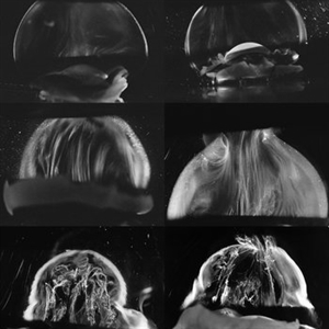
Dark-field slit microscopy was performed on fresh, unfixed, post-mortem human eyes that had undergone dissection to peel off the sclera, choroid, and retina. The vitreous body remains attached to the anterior segment which is seen below, while the posterior pole is above in these images. The top panel demonstrates the absence of internal vitreous structures that scatter light in youth (left image from an 11 year-old girl, right image from a 14 year-old boy. The middle panel demonstrates light scattering from linear, fibrous structures that have an antero-posterior orientation with insertions into the vitreous base peripherally and the posterior vitreous cortex, typical in middle age (left image from a 56 year-old and right image from a 59 year-old). The bottom panel illustrates advance fibrous liquefaction in old age (88-year-old subject). [From Sebag J, Niemeyer M, Koss M: Anomalous PVD and vitreoschisis. In: Vitreous – in Health & Disease (J. Sebag, ed.) Springer, New York, 2014, pg. 245; image © Springer Nature, reprinted with permission]
Condition/keywords: vitreous
-
 Aphakic Retinal Detachment
Nov 9 2012 by Norman Byer
This is an aphakic retinal detachment in a 66-year-old woman. Note the prominent yellow line which probably represents the posterior border of the vitreous base. Near the left end of the line there is a small retinal flap. At the exact location of this flap, there is a sudden break in the continuity of the yellow line, which is an important clue in finding this retinal tear. Often in aphakic detachments the causative tear is smaller than the one shown here and can be identified only by seeing a filamentous strand interrupting the uniform yellow line of the vitreous base.
Condition/keywords: aphakic retinal detachment, retinal flap, small retinal flap, vitreous base
-
 Avulsed Vitreous Base
-
 Encircling Buckle Effect
Jul 7 2015 by Hamid Ahmadieh, MD
Late FA image of the right eye of a 30-year-old man who underwent pars plana vitrectomy , endolaser photocoagulation and an encircling band placement a couple of years before following a penetrating trauma at the vitreous base area at the 7 o'clock meridian.
Photographer: Nayereh Hadipour, Negah Eye Center,Tehran, Iran
Imaging device: Specteralis
Condition/keywords: pars plana vitrectomy (PPV)
-
---thumb.jpg/image-square;max$300,300.ImageHandler) Floaters
Floaters
Oct 9 2013 by Maurice F. Rabb
KR is a 25 year old white female who presented with a one month history of floaters OD. Past ocular and systemic history were unremarkable. On clinical examination, the visual acuity was 20/20 OU, and the anterior segments were normal. There was a very mild degree of vitreous cell OD, though no cystoid macular edema nor vasculitis. A lobulated white mass was noted overlying the vitreous base inferotemporally OD (thickness 3.3mm). There was no calcification, though prominent cysts were noted on the surface of the lesion. A fluorescein angiogram, echogram, and CT scan were obtained, along with a thorough systemic evaluation.
Condition/keywords: floaters
-
 Human Vitreous Base Structure
Sep 1 2020 by J. Sebag, MD, FACS, FRCOphth, FARVO
Dark-field slit microscopy was performed on fresh, unfixed, post-mortem human eyes that had undergone dissection to peel off the sclera, choroid, and retina. The vitreous body remains attached to the anterior segment which is seen below, while the posterior pole is above in these images. Left: specimen was tilted to reveal the posterior aspect of the lens (L) and the fibers of the vitreous base (arrow) splayed out to insert anterior and posterior to the ora serrata; Right: Anterior Loop of the vitreous base (see text). [From Sebag J: The Vitreous - Structure, Function, and Pathobiology. Springer-Verlag, New York, 1989, pp. 41 & 42; images © Springer Nature, reprinted with permission]
Condition/keywords: vitreous
-
 IOFB
IOFB
Oct 11 2024 by Virginia Gebhart
55 year old male s/p RD repair with SB and SO in Mexico June 2023. Questionable foreign body inferior vitreous base. Pt asymptomatic, had no previous knowledge of IOFB.
Photographer: Virginia Gebhart, Retina Consultants of Carolina
Imaging device: Optos California
Condition/keywords: intraocular foreign body, IOFB, scleral buckle
-
 Peripheral Retinal Lesion
Nov 9 2012 by Norman Byer
This small elevated peripheral retinal lesion in a 48-year-old woman is a cystic retinal tuft. Such tufts are congenital developmental anomalies present from birth and situated behind the vitreous base. They are sites of abnormal vitreoretinal attachment, and can occasionally lead to retinal tears at the time of posterior vitreous detachment. They are present in about 5% of patients.
Condition/keywords: abnormal vitreal retinal attachment, behind the vitreous base, congenital anomaly, cystic retinal tuft, developmental anomaly, peripheral retinal lesion, present from birth
-
 Retinal Detachment
Nov 9 2012 by Norman Byer
This is a retinal detachment in a 55-year-old man. The vertical convex line on the right side probably represents the posterior border of the vitreous base. Note the small tractional tear with the base of its flap attached at this line. This demonstrates how the vitreous base presents an effective barrier to further extension of the retinal tear. Note also how the flap breaks the continuity of the yellow line.
Condition/keywords: retinal degeneration, retinal flap, tractional retinal tear, vitreous base
-
 Retinal Dialysis
Jul 5 2025 by Gustavo Uriel Fonseca Aguirre
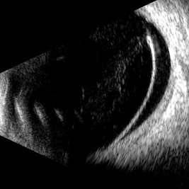
This B-mode longitudinal ultrasound scan demonstrates a retinal dialysis, appearing as a linear discontinuity at the ora serrata with associated vitreous base avulsion. The scan reveals mild subretinal fluid extending from the dialysis site with macular involvement.
Photographer: Gustavo U. Fonseca Aguirre, Hospital Conde de Valenciana, Ciudad de México
Condition/keywords: retinal dialysis
-
 Slide 8-19
Mar 4 2019 by Lancaster Course in Ophthalmology
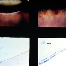
Traumatic retinal dialysis in a 15-year-old male who died 2 weeks following a motorcycle accident. Upper left shows dialysis of the retina at the ora serrata. Upper right shows hemorrhage In the retina of the fellow eye. Sections through dialysis (lower views) show the detached vitreous base with a small tag of adherent retinal tissue (arrow). (E.P. No. 32955)
Condition/keywords: hemorrhage, ora serrata, retinal dialysis
-
 Vitreous Base Avulsion
Feb 21 2022 by Maxwell J Wingelaar, MD
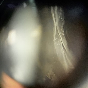
24-year-old male with a vitreous base avulsion with a history of blunt force trauma to the eye
Condition/keywords: vitreous detachment
-
 Vitreous Base Avulsion
Feb 21 2022 by Maxwell J Wingelaar, MD
24-year-old male with a vitreous base avulsion with a history of blunt force trauma to the eye
Condition/keywords: vitreous detachment
-
 Vitreous Body
Jan 3 2020 by Manuel Ángel Alcántara Delgado, MD
Slit lamp photograph of a 35-year-old woman with history of floaters.
Photographer: Manuel Ángel Alcántara Delgado, CMN SXXI, Mexico City
Condition/keywords: vitreous, vitreous base, vitreous floaters
 Loading…