Search results (22 results)
-
 Proliferative diabetic retinopathy
Jul 15 2022 by Rinat Sutiushev
Proliferative diabetic retinopathy with vitreoretinal traction and traction retinal detachment.
Photographer: Rinat Sutiushev
Condition/keywords: proliferative diabetic retinopathy (PDR)
-
 ROP
ROP
Mar 26 2025 by Korey Starkey
9 month old patient presents today with Retinopathy of Prematurity in both eyes. Patient was born at gestational age of 25 weeks 2 days, 940g. Left eye presents with vitreoretinal traction and peripheral VH with regressed stage 3 and persistent stage 2 disease.
Photographer: Korey Starkey
Imaging device: Optos
Condition/keywords: retinopathy of prematurity stage 2, rop, stage 3, vitreoretinal traction, vitreous hemorrhage
-
 Retinal Break at Site of Lattice Degeneration with Scleral Indentation
Nov 9 2012 by Norman Byer
This is the same case as the previous photograph. With scleral indentation slightly more posterior, the flap is seen to be associated with a large retinal tear. This is a tractional tear and it is possible that in this case the cryotherapy itself may have increased the vitreoretinal traction at this site and in this way led to this new tear. The age of the tear is unknown because it was asymptomatic, and even though the eye is aphakic the tear has not caused a clinical retinal detachment.
Condition/keywords: retinal flap, scleral indentation, tractional retinal tear, vitreoretinal traction
-
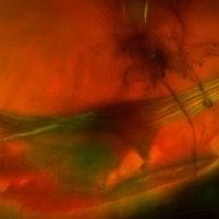
 Toxoplasmosis Vitreoretinal Traction
Jun 30 2013 by Jason S. Calhoun
A 28-year old male who noticed an upper partial visual loss to his right eye for about 2 weeks. Diagnosed with retinitis and was put on acyclovir. Patient received second opinion due to culture of aqueous humor which was then diagnosed as toxoplasmosis. VA was 20/25 in the right eye. Patient has been followed after for the past 7 months.
Photographer: Jason S. Calhoun, Mayo Clinic Jacksonville, Florida
Condition/keywords: toxoplasmosis
-
---thumb.JPG/image-square;max$300,300.ImageHandler) Toxoplasmosis Vitreoretinal Traction
Toxoplasmosis Vitreoretinal Traction
Jun 30 2013 by Jason S. Calhoun
A 28-year old male who noticed an upper partial visual loss to his right eye for about 2 weeks. Diagnosed with retinitis and was put on acyclovir. Patient received second opinion due to culture of aqueous humor which was then diagnosed as toxoplasmosis. VA was 20/25 in the right eye. Patient has been followed after for the past 7 months.
Photographer: Jason S. Calhoun, Mayo Clinic Jacksonville, Florida
Condition/keywords: toxoplasmosis
-
 Asymptomatic Superior Retinal Detachment
May 5 2016 by Steven J Ryder, MD
38-year-old African American female with moderate myopia (-4.50 Sph OU) and asymptomatic superior retinal detachment in the right eye. Zeiss Cirrus OCT capturing full-thickness retinal break at 12:00 and temporal vitreoretinal traction.
Photographer: Luis Bernhard, Miami VA Healthcare System
Imaging device: Zeiss Cirrus
Condition/keywords: asymptomatic, full thickness retinal hole, retinal break, retinal detachment with retinal defect
-
 Asymptomatic Tractional Tear
Nov 9 2012 by Norman Byer
This 38-year-old man was found to have this asymptomatic tractional tear in which the vitreoretinal traction had completely avulsed this tiny fragment of retina as a free operculum. Note how the examination and also the photography of this tiny lesion is made easier by scleral indentation.
Condition/keywords: asymptomatic, free operculum, scleral indentation, vitreoretinal traction
-
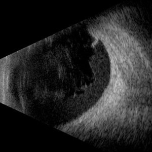 Hemorrhagic Vitreous Detachment
May 21 2025 by Gustavo Uriel Fonseca Aguirre
This B-mode longitudinal ultrasound scan shows a hemorrhagic vitreous detachment with the peripheral hyaloid strongly adherent to a retinal break. Associated vitreous and subhyaloid hemorrhage are present, indicating acute vitreoretinal traction.
Photographer: Gustavo U. Fonseca Aguirre, Hospital Conde de Valenciana, Ciudad de México
Condition/keywords: Hemorrhagic Vitreous Detachment
-
 Laser Photocoagulation
Nov 9 2012 by Norman Byer
This is the same lesion 18 days following photocoagulation. The continuing vitreoretinal traction has now torn the retinal flap completely away from the retina and the resulting free operculum may be seen out of focus in the lower part of the photograph. The retinal tear is now easily visible with only a tiny remaining nubbin of the original flap seen above with a small hemorrhage.
Condition/keywords: free operculum, laser photocoagulation, retinal tear, vitreoretinal traction
-
 Lattice Degeneration
Nov 9 2012 by Norman Byer
In this 54-year-old woman, lattice degeneration has led to a large horseshoe tractional tear around the posterior side on one end of the lesion resulting in a clinical retinal detachment. Note the very attenuated blood column passing through the white sheath vessel that crosses the tear. This demonstrates that the white blood vessels and a fragment of attached tissue are the only structures which have escaped the tearing effect of the strong vitreoretinal traction which occurred. This usually is true, although, in some cases this bridging vessel may bleed.
Condition/keywords: bridging vessel, lattice degeneration, tractional retinal tear, white sheath vessel
-
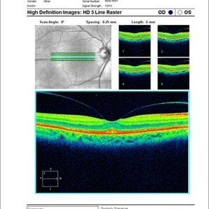
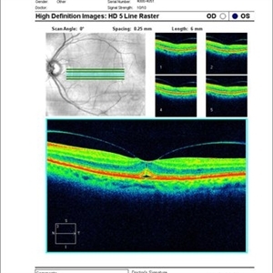
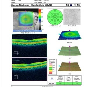
 Macular Vitreotraction
Aug 25 2017 by JEFFERSON R SOUSA, Tecg.º (Biomedical Systems Technology)
A 56-year-old female patient had a low vision clinic, a vitreoretinal traction was observed on the OCT examination.
Photographer: JEFFERSON R SOUSA - Study Center and Ophthalmological Research Dr. Andre M V Gomes, Dr. Suel Abujamra Institute São Paulo-Brazil
Imaging device: OCT CIRRUS 4000, Protocol Line 6mm, 90 degrees.
Condition/keywords: macular traction
-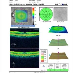
 Sudden Posterior Vitreous Detachment
Nov 9 2012 by Norman Byer
This is the appearance of the previous lesion three weeks following prophylactic cryotherapy. Continuing vitreal retinal traction has a now torn the flap completely free from the retina. The whitish cystic retinal tuft can be discerned on the upper part of the free operculum. Along the lower half of the operculum superimposed over the dark shadow of the scleral indentation one may observe numerous, delicate, vitreous fibrils actually attaching to the operculum.
Condition/keywords: cystic retinal tuft, free operculum, prophylactic cyrotherapy, retinal flap, scleral indentation, vitreoretinal traction, vitreous fibrils
-
 Vascular Loop
Mar 26 2019 by Gary R. Cook, MD, FACS
53-year-old diabetic African American male with a venous vascular loop secondary to focal vitreoretinal traction on the vessel; healed laser PRP scars visible; VA = 20/200
Imaging device: Topcon VT-50
Condition/keywords: vascular loop
-
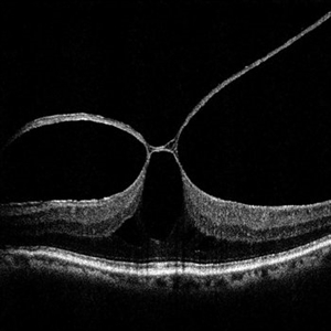
 Vitreal Macular Traction
Mar 27 2013 by David W. Faber, MD
Vitreal Macular Traction.
Condition/keywords: vitreoretinal traction
-
 Vitreal Macular Traction
Mar 27 2013 by David W. Faber, MD
Vitreal Macular Traction.
Condition/keywords: vitreoretinal traction
-
 Vitreal Macular Traction
Mar 27 2013 by David W. Faber, MD
Vitreal Macular Traction.
Condition/keywords: vitreoretinal traction
-
 Vitreal Macular Traction
Mar 27 2013 by David W. Faber, MD
Vitreal Macular Traction.
Condition/keywords: vitreoretinal traction
-
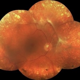
 Vitreoretinal Traction with Adjacent Tear and Vitreous Hemorrhage
Oct 3 2023 by Alexis Singstock
Ultra-widefield fundus photograph of a 76 year old woman with vitreoretinal traction, an adjacent retinal tear and vitreous hemorrhage affecting the left eye. Patient was referred for retinal detachment and vitreous hemorrhage. Patient reports waking up the day prior to their appointment with "a lot of lines coming down the front, like swirling dirt in the left eye". Patient's vision was counting fingers at 1 ft. Dr. Joseph Boss noticed a horseshoe tear inferior to traction on exam and with the help of ultra-widefield imaging. Dr. Boss performed laser retinopexy to tear and impending tear at site of traction. Patient is scheduled for pars plana vitrectomy for dense vitreous hemorrhage.
Photographer: Alexis Singstock
Imaging device: Optos California
Condition/keywords: acute posterior vitreous detachment, fundus photography, left eye, Optos, OPTOS CALIFORNIA, pseudocolor, ULTRA WIDE FIELD, vitreoretinal traction, vitreous hemorrhage
-
 White Without Pressure and Peripheral Retinoschisis
Dec 29 2022 by Gulnara Islamova
Fundus Photograph and OCT scan of an 18 year-old male with peripheral retinoschisis combined with WWOP lessions .Vitreoretinal traction is not visualized
Photographer: Gulnara Islamova, CENTER ZRENIYA Medical Clinic, LLC, Chelyabinsk, Russian Federation
Imaging device: Optovue XR Avanti
Condition/keywords: peripheral retinal degeneration
 Loading…