Search results (2 results)
-
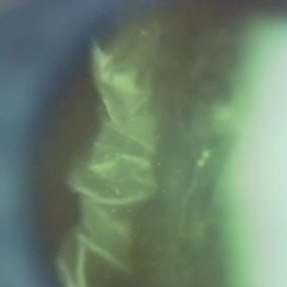 Folds in Detached Posterior Vitreous Cortex
May 31 2022 by Joshua Friedman
Slit lamp (video) image showing folds in the posterior vitreous cortex in an eye with PVD.
Photographer: Martin Snead, MD, Cambridge, England
Condition/keywords: folds, posterior vitreous cortex, PVD, vision degrading myodesopsia, vitreous
-
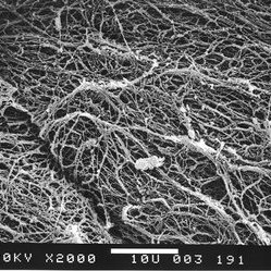 Scanning electron microscopy of the posterior aspect of the human posterior vitreous cortex
May 31 2022 by Joshua Friedman
Scanning electron microscopy demonstrates the dense packing of collagen fibrils in the posterior vitreous cortex. To some extent this arrangement is exaggerated by the dehydration that occurs during specimen preparation for scanning electron microscopy (bar = 10 µm).
Photographer: EM lab, Eye Research Institute of Retina Foundation, Boston, MA
Condition/keywords: collagen fibrils, posterior vitreous cortex, scanning EM, vision degrading myodesopsia
 Loading…