Search results (2 results)
-
 Penetrating Trauma of an Inadvertent Sub-Tenon's Kenalog Injection
Jan 31 2018 by Olivia Rainey
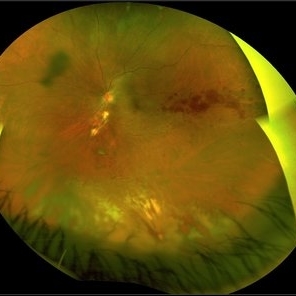
Ultra-wide field pseudocolor photograph of a 38-year-old female with penetrating trauma after an inadvertent sub-tenon's kenalog injection affecting her left eye. Patient has a large dehemoglobinized vitreous hemorrhage settling inferior near the entry wound. The exit wound has developed chorioretinal scarring and the disruption of several veins near the optic nerve, resulting in a branch retinal vein occlusion.
Photographer: Olivia Rainey
Imaging device: Optos
Condition/keywords: branch retinal vein occlusion (BRVO), chorioretinal scar, color fundus photograph, dehemoglobinized hemorrhage, kenalog, left eye, montage, Optos, penetrating trauma, sub-tenon's, ultra-wide field imaging
-
 Traumatic Retinal Detachment
May 5 2025 by Gustavo Uriel Fonseca Aguirre
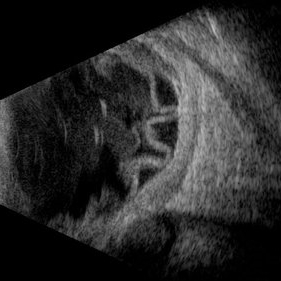
This B-mode longitudinal ultrasound scan over the macular area reveals vitreous hemorrhage, retinal detachment with folding, peripheral annular choroidal detachment, and sub-Tenon's fluid in the setting of blunt ocular trauma. The findings indicate severe posterior segment disruption with multi-compartment involvement.
Photographer: Gustavo U. Fonseca Aguirre, Hospital Conde de Valenciana, Ciudad de México
Condition/keywords: blunt trauma, Retinal Detachment
 Loading…