Search results (8 results)
-
 Asymptomatic Lesion
Nov 9 2012 by Norman Byer
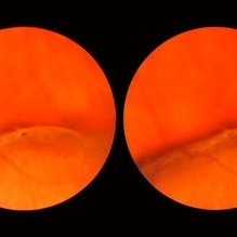
This is the same lesion as seen in the previous slide pair. Here the scleral indentation is carried more posterior revealing a tiny, round, full thickness retinal hole. This is not a tear produced by traction even though vitreous is always attached to these flaps. You will note that the hole is round and is separated by a slight distance from the flap itself. It is probably the result of long continued atrophy and devitalization of the retina. A posterior vitreous was not detached. This lesion has not changed its appearance for more than a year of observation, but the age of the hole is actually unknown.
Condition/keywords: asymptomatic, atrophy, full thickness retinal hole, posterior scleral indentation, retinal hole, round hole
-
 Flat Lattice Lesion
Nov 9 2012 by Norman Byer
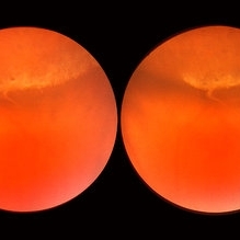
This 34 year-old man had a flat lattice lesion with no hole at this location for five years. Then he developed this round hole with a small subclinical retinal detachment which has not changed in appearance for four years. Note the tiny glial tuft just to the left of the hole and superimposed against the dark background.
Condition/keywords: glial tuft, lattice degeneration, round hole
-
 Lattice Combined with Tiny Round Hole
Nov 9 2012 by Norman Byer
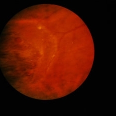
This 45 year-old man shows the snail track form of lattice combined with a tiny round hole. There is a tiny subclinical retinal detachment confined to the lesion itself.
Condition/keywords: glial vitreous tuft, lattice degeneration, round hole, snail track
-
 Lattice Degeneration
Jan 5 2015 by H. Michael Lambert, MD
Lattice degeneration with round holes.
Condition/keywords: lattice degeneration
-
 Lattice Lesion
Nov 9 2012 by Norman Byer
When this boy was first examined at the age of six years, he had only the red crater form of lattice at this location. This photograph shows the same lesion at age 11 and there is now a small round atrophic hole with a tiny round zone of detachment around it. It has not changed for four years.
Condition/keywords: atrophic retinal hole, lattice degeneration, lattice lesion, reddish crater, round hole
-
 Lattice Lesion
Nov 9 2012 by Norman Byer
This lattice lesion in a 44-year-old woman shows combined features of pigmentation, white lines, yellow dots and a round hole with a tiny zone of adjacent detachment. There are three such holes in this eye and they have not changed or been treated for eight years.
Condition/keywords: adjacent detachment, atrophic retinal hole, lattice degeneration, lattice lesion, pigmented lattice lesion, round hole, white lattice lines, yellow dots
-
 Slide 9-47
Feb 26 2019 by Lancaster Course in Ophthalmology
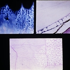
Typical peripheral cystoid degeneration. A small, round hole within the area of cystoid degeneration is present (upper left). In section the cysts are located mostly in the outer plexiform layer (upper left). A nondigestion flat preparation of the retina shows the cystic spaces, which have coalesced to form meridionally oriented tunnels (lower view).
Condition/keywords: peripheral cystoid degeneration
-
 Subclinical Retinal Detachment
Nov 9 2012 by Norman Byer
This 50-year-old man was treated with cryotherapy for two tiny non tractional round holes which had produced a small subclinical retinal detachment at 7:00 o’clock in this eye. Two years later he was seen with this large horseshoe tractional tear just anterior to the treated area and we must assume that it was a complication of that treatment.
Condition/keywords: cryotherapy, non-tractional holes, tractional retinal tear
 Loading…