Search results (453 results)
-
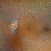
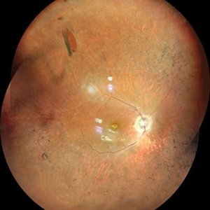
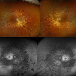
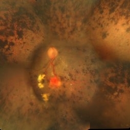 Retinitis Pigmentosa With Hemangioma CF
Dec 15 2016 by Manish Nagpal, MD, FRCS (UK), FASRS
Fluorescein angiography OS of a patient having retinitis pigmentosa with a hemangioma inferiorly.
Condition/keywords: hemangioma, retinitis pigmentosa
-
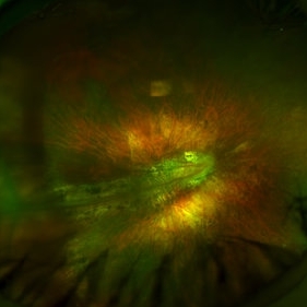
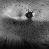 Phoenix
Feb 21 2024 by Sayena . Jabbehdari, MD, MPH, MBA
A 60-year-old Caucasian female presented with reduced night vision and constricted visual fields. The fundus exam revealed pigmentary changes in the peripheral retina. Fundus autofluorescence depicted the schematic appearance of a Phoenix , with the hypo-autofluorescence corresponding to the head and wings of the phoenix. Genetic testing was positive for a heterozygous RHO mutation
Photographer: Sayena Jabbehdari MD MPH
Condition/keywords: retinitis pigmentosa
-
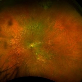
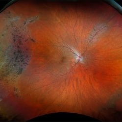
 Retinitis Pigmentosa with Macular Hole with Posterior Subcapsular Cataract
Apr 28 2025 by Malvika Singh
Fundus photograph of the right eye of a 31 year old with retinitis pigmentosa with a macular hole, showing the shadow of posterior subcapsular cataract over the fundus.
Photographer: Dr Malvika Singh, Retina Foundation, Ahmedabad, India
Imaging device: Mirante SLO/OCT
Condition/keywords: macular hole, posterior subcapsular cataract, retinitis pigmentosa
-
 Autofluorescence of Retinitis Pigmentosa
Jul 13 2016 by Linda A Cernichiaro- Espinosa, MD
Fundus autofluorescence of an 53-year-old woman with retinitis pigmentosa.
Photographer: Tec Ricardo Montoya, Clínica Oftalmológica Anzures
Condition/keywords: retinitis pigmentosa
-
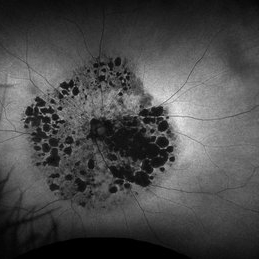
 Bone Corpuscle Pigments
Sep 11 2014 by Mehul A Shah
A 42-year-old female presented with gradual reduction in vision.
Photographer: Drashti Netralaya,Dahod
Imaging device: FF 450
Condition/keywords: retinitis pigmentosa (RP) dystrophy
-
 Optic Disc Drusen, RP
Apr 21 2025 by Virginia Gebhart
28 year old male with stable retinitis pigmentosa and optic disc drusen OU. Bardet-Biedl variant identified in previous genetic testing. BCVA 20/50 OD, 20/30 OS
Photographer: Virginia Gebhart, Retina Consultants of Carolina
Imaging device: Optos California
Condition/keywords: Drusen, optic disc drusen, retinitis pigmentosa
-
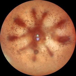
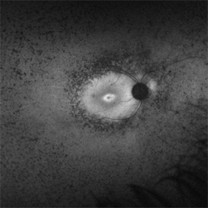
 Pericentral Retinitis Pigmentosa
Sep 6 2024 by Mauricio Bayram-Suverza, MD
A 65-year-old male patient reports experiencing bilateral blind spots that have gradually intensified over time. Genetic testing was unrevealing. The fundus autofluorescence image shows a hypoautofluorescent ring in the posterior pole, especially nasal to the nerve and along arcades.
Photographer: Mauricio Bayram-Suverza, Casey Eye Institute, OHSU.
Imaging device: Optos California
Condition/keywords: fundus autofluorescence (FAF), inherited retinal disease, nyctalopia, retinal dystrophy, retinitis pigmentosa
-
 Retinitis Pigmentosa with PPRPE
Jan 27 2025 by Vishal Agrawal, MD, FRCS,FACS,FASRS
16 year-old male patient presented with DOV, nyctalopia and nystagmus. Fundus revealed pigment clumping, pale disc and preserved para-arteriolar retinal pigment epithelium (PPRPE) in both eyes. Genetic testing revealed CRB1 gene mutation.
Photographer: Dr Ayushi
Imaging device: Clarus 700
Condition/keywords: retinitis pigmentosa
-
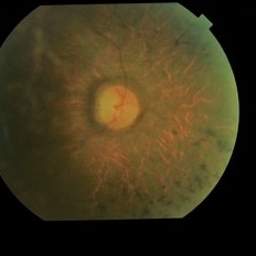
 Advanced Retinitis Pigmentosa
Mar 29 2024 by Aditya S Kelkar, MS, FRCS, FASRS,FRCOphth
Fundus photograph of an 70-year-old man with advanced retinitis pigmentosa.
Photographer: Optom Ayesha Inamdar, National Institute of Ophthalmology, Pune
Imaging device: Daytona OPTOS
Condition/keywords: RETINITIS PIGMENTOSA, RP
-
 Case 2 Retinitis Pigmentosa BAF IRAF OD
May 14 2014 by Avris Romario Diparaja Siahaan
Fundus image a 57-year-old man with retinitis pigmentosa on both eyes. These image were taken with blue auto fluorescein mode (BAF) and infrared auto fluorescence (IRAF).
Photographer: Avris Romario Diparaja Siahaan
Imaging device: Heidelberg HRA + OCT Spectralis
Condition/keywords: autofluorescence imaging, fundus photograph, infrared image, retinitis pigmentosa
-
 Case 2 Retinitis Pigmentosa BAF IRAF OS
May 14 2014 by Avris Romario Diparaja Siahaan
Fundus image a 57-year-old man with retinitis pigmentosa on both eyes. These image were taken with blue auto fluorescein mode (BAF) and infrared auto fluorescence (IRAF).
Photographer: Avris Romario Diparaja Siahaan
Imaging device: Heidelberg HRA + OCT Spectralis
Condition/keywords: autofluorescence imaging, fundus photograph, infrared image, retinitis pigmentosa
-
 Dislocated Lens
Apr 26 2023 by Chloe Hanifan
Ultra wide field fundus photograph of a 41-year-old male with a dislocated lens affecting his right eye. IOL noted inferior vitreous base and vitrectomy surgery for removal of IOL was recommended. Patient has history of retinitis pigmentosa as well. Patient's vision at the time of presentation was counting fingers at 2 feet.
Photographer: Chloe Hanifan
Imaging device: Optos California
Condition/keywords: dislocated lens, fundus photography, Optos, pseudocolor, retinitis pigmentosa, ULTRA WIDE FIELD
-
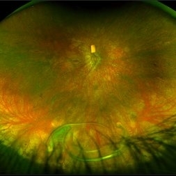
 Horseshoe Tear in Retinitis Pigmentosa
Mar 22 2021 by ASRS Staff
Montage of 25-year-old patient, high myopic patient came with complaint of diminution of vision in both eyes and on posterior segment examination of right eye, HST was present along with maculopathy.
Imaging device: Nidek Mirante
Condition/keywords: maculopathy, retinitis pigmentosa
-
 Mucopolysaccharidosis Type III
Apr 21 2023 by Matthew Dombrow, MD
Fundus photograph and autofluorescence of a 49 year old male with mucopolysaccharidosis type III (Sanfilippo syndrome)
Photographer: Cori Sturtevant, Connecticut Retina Consultants, Hamden, Connecticut
Condition/keywords: mucopolysaccharidoses, retinitis pigmentosa (RP) dystrophy
-
 Mucopolysaccharidosis Type III (Sanfilippo syndrome)
Apr 21 2023 by Matthew Dombrow, MD
Fundus photograph and autofluorescence of a 49 year old male with mucopolysaccharidosis type III (Sanfilippo syndrome)
Photographer: Cori Sturtevant, Connecticut Retina Consultants, Hamden, Connecticut
Condition/keywords: mucopolysaccharidoses, retinitis pigmentosa (RP) dystrophy
-
---thumb.jpg/image-square;max$300,300.ImageHandler) Retinitis Pigmentosa
Retinitis Pigmentosa
Oct 13 2012 by Geoffrey G. Emerson, MD, PhD, FASRS
Condition/keywords: bone spicule, retinitis pigmentosa
-
 Retinitis Pigmentosa
Mar 13 2013 by Carl C. Awh, MD, FASRS
Retinitis Pigmentosa
Photographer: Alecia Camp, CRA - Tennessee Retina - Nashville, TN
Condition/keywords: retinitis pigmentosa
-
 Retinitis Pigmentosa
Jul 12 2021 by Jeffrey Barker
FA OD
Photographer: Jeffrey P. Barker
Condition/keywords: retinitis pigmentosa
-
 Retinitis Pigmentosa
Jul 12 2021 by Jeffrey Barker
FA OS
Photographer: Jeffrey P. Barker, Retina Vitreous Surgeons of CNY.
Condition/keywords: retinitis pigmentosa
-
 Retinitis Pigmentosa
Apr 17 2025 by Virginia Gebhart
Fundus autofluorescence of 75 year old female with Retinitis Pigmentosa. Pt diagnosed at age 53. Diffuse RPE atrophy with minimal central sparing present in both eyes. Stable and unchanged compared to previous exams. BCVA 20/200 OD, NLP OS
Photographer: Virginia Gebhart, Retina Consultants of Carolina
Imaging device: Optos California
Condition/keywords: autofluorescence imaging, bone spicule, retinitis pigmentosa, RP
-
 Retinitis Pigmentosa
Apr 9 2025 by Virginia Gebhart
35 year old female with stable sectoral RP and high myopia OU. RP has not progressed in either eye since initial visit in 2021. Will continue to observe. VA 20/20 OU
Photographer: Virginia Gebhart, Retina Consultants of Carolina
Imaging device: Optos California
Condition/keywords: high myopia, retinitis pigmentosa
-
 Retinitis Pigmentosa
Nov 7 2023 by Jolee Rodriguez
Bilateral fundus photography and fundus autofluorescence imaging of a 62-year-old male with Retinitis Pigmentosa. Patient reported visual field defects and dark adapting issues. Patient's vision at the time images were taken were sc20/20 of the right eye and sc20/25 of the left eye. Dr. Sutherland determined that based on the patient's lack of family history, the most likely route of inheritance is autosomal recessive.
Photographer: Jolee Rodriguez
Imaging device: Optos California RGB
Condition/keywords: autofluorescence imaging, fundus photography, hereditary retinal dystrophy, Optos, OPTOS CALIFORNIA RGB, retinitis pigmentosa, ultra-wide field imaging, Ultra-wide field retinal imaging, ultra-widefield image
-
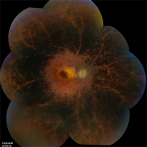
 Retinitis Pigmentosa and Foveal Dragging
Dec 30 2012 by Barbara Parolini, MD
Panoramic fundus photograph of the right eye of a 37-year-old woman with retinitis pigments and foveal dragging. BCVA is 20\200
Photographer: Barbara Parolini, MD
Imaging device: Daytona
Condition/keywords: retinitis pigmentosa
-
 Retinitis Pigmentosa Bullseye Appearing Autofluorescence
Feb 4 2025 by Isaac Agranoff
Fundus Autofluorescence of a 14-year-old boy with suspected RP. ERG performed afterwards was almost flat. VA measured at 20/30 but with extensive constriction of confrontational visual fields. Currently awaiting genetic testing.
Photographer: Isaac Agranoff
Imaging device: Optos California
Condition/keywords: fundus autofluorescence (FAF), retinitis pigmentosa, RP
-
 Retinitis Pigmentosa in Laurence Moon Biedl Bardet Syndrome
Oct 19 2017 by yashaswi pendyala
Fundus photograph of an 32-year-old male with features suggestive of Laurence Moon Biedl Bardet Syndrome.
Photographer: yashaswi pendyala ,mamata general hospital ,department of ophthalmology
Condition/keywords: retinitis pigmentosa
 Loading…