Search results (34 results)
-
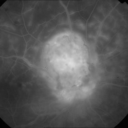
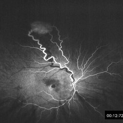
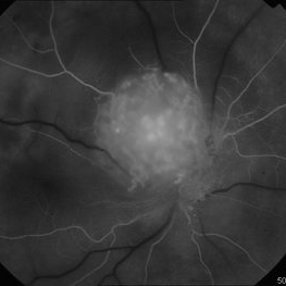 Early Phase FA of Optic Disc Capillary Hemangioblastoma
Mar 18 2014 by Arwa Azmeh, MD, PhD
FA showed early hyperfluorescent spots over the mass.
Condition/keywords: retinal hemangioblastoma
-
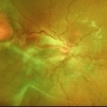
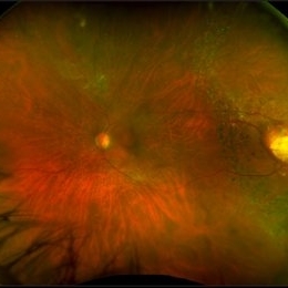 Hemangioma
Feb 9 2021 by Kim Barrett
66-year-old female with a history of thyroid and uterine cancer in her 30's. She has a family history of cancers also. Current VA 20/40-2 PH OS. Patient and doctor chose observation at this time with possible surgical intervention in the future. She also has a small Hemangioma temporally in the right eye. Von Hippel-Lindau is also suspected and genetic testing was suggested.
Photographer: Kim Barrett C.O.A. Retina Specialists of Michigan, Grand Rapids, MI
Imaging device: Optos California
Condition/keywords: cancer, genetic testing, Optos, retinal hemangioblastoma, Von Hippel-Lindau
-
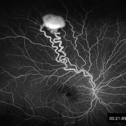 Retinal Hemangioblastoma Mid Phase FA
May 15 2013 by Robert T. Wendel, MD
20-year-old male. Genetic hx not yet defined.
Condition/keywords: Von Hippel-Lindau
-
 Solitary Capillary Retinal Hemangioblastoma
Apr 29 2019 by Michael A. Novak, MD
17-year-old young lady presented with reduced vision OD for several months. Her vision was 20/50.
Condition/keywords: retinal capillary hemangioblastoma, Von Hippel-Lindau
-
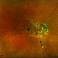
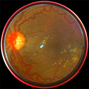
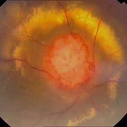 Color Photo of Optic Disc Capillary Hemangioblastoma
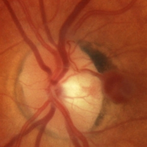
Mar 18 2014 by Arwa Azmeh, MD, PhD
Color fundus photograph of an 48-year-old male who complained of decreased visual acuity in his right eye over the last few months. Systemically the patient was healthy. His VA was OD Cf 3m, OS 20/20. Anterior segments were WNL in OU. IOP was WNL in OU. Fundus exam OD revealed unpigmented mass over the optic disc with retinal venous tortuosity at its edges with a ring of thick HYE surrounding it and shallow RD in this area extending to the foveal area. Several few small retinal hemorrhages were seen in the far retinal periphery which were explained to be caused by venous stasis due the optic disc tumor.
Condition/keywords: color photo, optic disc, retinal hemangioblastoma
-
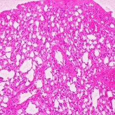 Gross Pathology of Retinal Hemangioblastoma
Nov 20 2019 by McGill University Health Centre
39-year-old Caucasian man with a diagnosis of Von Hippel-Lindau Disease. Eyes were obtained post-mortem. Gross pathology: hemangioblastomas of the retina are represented by vascular proliferation.
Condition/keywords: retinal hemangioblastoma, Von Hippel-Lindau
-
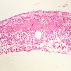 Gross Pathology of Retinal Hemangioblastoma
Nov 20 2019 by McGill University Health Centre
39-year-old Caucasian man with a diagnosis of Von Hippel-Lindau disease. Eyes were obtained post-mortem. Gross pathology: hemangioblastomas of the retina are represented by vascular proliferation.
Condition/keywords: retinal hemangioblastoma, Von Hippel-Lindau
-
 Hemangioblastoma
Sep 15 2017 by Jason Griffith
17-year-old female with family history of renal cell carcinoma and sibling with cerebellar hemangioblastoma. Patient sent for MRI study to rule out Von Hippel Lindau Syndrome.
Photographer: Jason Griffith, Tennessee Retina, Nashville, TN
Imaging device: Optos California
Condition/keywords: retinal hemangioblastoma, Von Hippel-Lindau
-
 Hemangioblastoma Post PDT X2
-
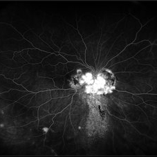
 Late Phase FA of Optic Disc Capillary Hemangioblastoma
Mar 18 2014 by Arwa Azmeh, MD, PhD
Late phase FA showed increased hyper fluorescence of the mass.
Condition/keywords: optic disc, retinal hemangioblastoma
-
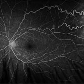
 Optic Disc Hemangioblastoma
May 30 2017 by Olivia Rainey
Ultra-wide-field fluorescein angiogram of the right eye of an 29-year-old female with an optic nerve hemangioblastoma secondary to Von Hippel-Lindau Syndrome.
Photographer: Olivia Rainey
Imaging device: Optos California
Condition/keywords: fluorescein angiogram (FA), fluorescein leakage, optic disc, Optos, retinal hemangioblastoma, ultra-wide field imaging, Von Hippel-Lindau
-
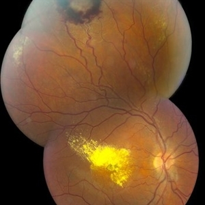
 Optic Nerve Hemangioblastoma
May 30 2017 by Olivia Rainey
Ultra-wide-field color fundus photograph of the right eye of an 29-year-old female with an optic nerve hemangioblastoma secondary to Von Hippel-Lindau Syndrome.
Photographer: Olivia Rainey
Imaging device: Optos California
Condition/keywords: color fundus photograph, optic nerve, Optos, retinal hemangioblastoma, ultra-wide field imaging, Von Hippel-Lindau
-
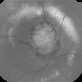 Red Free Photo of Optic Disc Capillary Hemangioblastoma
Mar 18 2014 by Arwa Azmeh, MD, PhD
Red free fundus photograph of an 48-year-old male who complained of decreased visual acuity in his right eye over the last few months. Systemically the patient was healthy. His VA was OD Cf 3m, OS 20/20. Anterior segments were WNL in OU. IOP was WNL in OU. Fundus exam OD revealed unpigmented mass over the optic disc with retinal venous tortuosity at its edges with a ring of thick HYE surrounding it and shallow RD in this area extending to the foveal area. Several few small retinal hemorrhages were seen in the far retinal periphery which were explained to be caused by venous stasis due to the optic disc tumor
Condition/keywords: optic disc, red-free, retinal hemangioblastoma
-
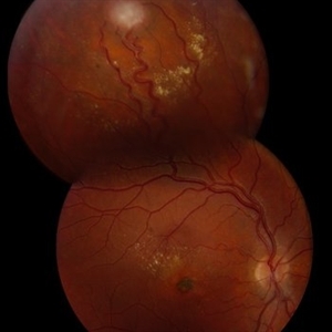
---thumb.jpg/image-square;max$300,300.ImageHandler) Retinal Hemangioblastoma
Retinal Hemangioblastoma
Feb 20 2013 by From the Collections of Thomas M. Aaberg, MD and Thomas M. Aaberg Jr., MD
Feeder vessels to retinal mass
Condition/keywords: retinal hemangioma
-
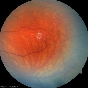
 Retinal Hemangioblastoma
Oct 16 2019 by Prithvi Chandrakanth
30-year-old male, fundus examination revealed retinal hemangioblastoma in the inferotemporal quadrant.
Photographer: Dr.Prithvi Chandrakanth, Dr.Chandrakanth Malabar Nethralaya, Kozhikode, India
Imaging device: TRASH TO TREASURE RETCAM
Condition/keywords: retcam, retinal hemangioblastoma, smartphone fundus photography, Von Hippel-Lindau
-
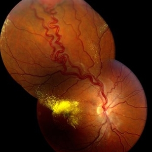
 Retinal Hemangioblastoma
Aug 10 2024 by Muhammad Amer Awan, MD, FRCSEd, FRCOphth, FRCS Glasgow, FACS, FASRS
Fundal photograph of a 21 years old female with a large and small retinal hemangioblastoma with extensive epiretinal membrane over the macula and macular detachment in the right eye.
Condition/keywords: retinal hemangioblastoma
-
 Retinal Hemangioblastoma
May 15 2013 by Robert T. Wendel, MD
20-year-old male. Genetic hx not yet defined.
Condition/keywords: Von Hippel-Lindau
-
 Retinal Hemangioblastoma
Jun 30 2014 by Robert T. Wendel, MD
Retinal hemangioblastoma.
Condition/keywords: retinal hemangioblastoma
-
 Retinal Hemangioblastoma
Jun 30 2014 by Robert T. Wendel, MD
Retinal hemangioblastoma.
Condition/keywords: retinal hemangioblastoma
-
 Retinal Hemangioblastoma early FA
May 15 2013 by Robert T. Wendel, MD
20-year-old male. Genetic hx not yet defined.
Condition/keywords: Von Hippel-Lindau
-
 Retinal Hemangioblastoma PO PDT
Jun 12 2013 by Robert T. Wendel, MD
Retinal hemangioblastoma, 10 days post full fluence PDT.
-
 Same Patient
Jul 1 2014 by John S. King, MD
Same patient, a different lesion: Small retinal hemangioblastoma, laser performed around lesion to feeding arterioles, avoiding the drainage vein and body of lesion.
Photographer: Wayne A Ladlee Jr
Condition/keywords: retinal hemangioblastoma, Von Hippel-Lindau
-
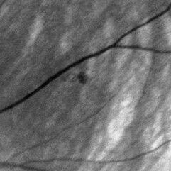 Same Patient
Jul 1 2014 by John S. King, MD
Same pateint, a different lesion: Very small lesion that appeared to be a very early hemangioblastoma, laser performed.
Photographer: Wayne A Ladlee Jr
Imaging device: Red Free
Condition/keywords: retinal hemangioblastoma, Von Hippel-Lindau
-
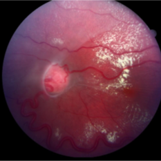
 VHL "Free Floating" Juxtapapillary Hemangioblastoma
Jul 1 2014 by John S. King, MD
30-year-old female with fhx VHL and CNS hemangioblastomas and visceral lesions. P/C with a floater (no PVD or VH) after episodes of vomiting.
Photographer: Wayne A Ladlee Jr
Condition/keywords: retinal hemangioblastoma, Von Hippel-Lindau
-
 VHL "Free Floating" Juxtapapillary Hemangioblastoma
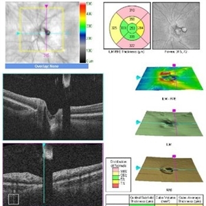
Jul 1 2014 by John S. King, MD
30-year-old female with fhx VHL and CNS hemangioblastomas and visceral lesions. P/C with a floater (no PVD or VH) after episodes of vomiting. - corresponds to earlier photos
Photographer: Wayne A Ladlee Jr
Imaging device: Cirrus
Condition/keywords: retinal hemangioblastoma, Von Hippel-Lindau
 Loading…