Search results (15 results)
-
 Retinal Break at Site of Lattice Degeneration with Scleral Indentation
Nov 9 2012 by Norman Byer
This is the same case as the previous photograph. With scleral indentation slightly more posterior, the flap is seen to be associated with a large retinal tear. This is a tractional tear and it is possible that in this case the cryotherapy itself may have increased the vitreoretinal traction at this site and in this way led to this new tear. The age of the tear is unknown because it was asymptomatic, and even though the eye is aphakic the tear has not caused a clinical retinal detachment.
Condition/keywords: retinal flap, scleral indentation, tractional retinal tear, vitreoretinal traction
-
 Acute Posterior Vitreous Detachment
Nov 9 2012 by Norman Byer
This large and complicated retinal tear in a 51-year-old man resulted from an acute posterior vitreous detachment which concentrated its tractional forces around this area of lattice degeneration. Because of the powerful traction, there is an additional central tear splitting the large retinal flap and almost severing one of its arms. The traction was strong enough to completely rupture the blood vessel just to the left of the flap. Marking the ruptured peripheral end of the blood vessel is a yellow depigmented thrombus.
Condition/keywords: acute posterior vitreous detachment, depigmented thrombus, lattice degeneration, retinal tear, tractional retinal detachment
-
 Aphakic Retinal Detachment
Nov 9 2012 by Norman Byer
This is an aphakic retinal detachment in a 66-year-old woman. Note the prominent yellow line which probably represents the posterior border of the vitreous base. Near the left end of the line there is a small retinal flap. At the exact location of this flap, there is a sudden break in the continuity of the yellow line, which is an important clue in finding this retinal tear. Often in aphakic detachments the causative tear is smaller than the one shown here and can be identified only by seeing a filamentous strand interrupting the uniform yellow line of the vitreous base.
Condition/keywords: aphakic retinal detachment, retinal flap, small retinal flap, vitreous base
-
 Dialysis of Retina in Upper Nasal Quadrant
Nov 9 2012 by Norman Byer
This 20-year-old wrestler sustained a sharp powerful blow to his right eye from his opponent’s thumb. One hour later he saw hundreds of black specs in his vision and was found to have this dialysis of his retina in the upper nasal quadrant. Note the triangular piece of retina in the center that remained attached to the ora serrata causing the retinal flap to resemble a man’s flared shirt collar.
Condition/keywords: retinal dialysis, retinal tear, upper nasal quadrant
-
 Giant Retinal Tear
Mar 29 2014 by Min Kim, MD, PhD, MBA, FASRS
Wide field fundus photograph of a 25 year-old male shows giant retinal tear with inverted retinal flap.
Photographer: Young Duk Bae, Yonsei University, Gangnam Severance Hospital
Imaging device: Optomap
Condition/keywords: giant retinal tear
-
 Giant Retinal Tear
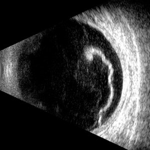
Jul 5 2025 by Gustavo Uriel Fonseca Aguirre
This B-mode longitudinal ultrasound scan reveals a giant retinal tear, demonstrating a circumferential retinal flap with rolled edges extending over M-X to M-I. The vitreous shows diffuse hemorrhage and anterior-posterior traction strands inserting at the tear margins. The remaining retina appears attached without subretinal fluid.
Photographer: Gustavo U. Fonseca Aguirre, Hospital Conde de Valenciana, Ciudad de México
Condition/keywords: giant retinal tear
-
 Laser Photocoagulation
Nov 9 2012 by Norman Byer
This is the same lesion 18 days following photocoagulation. The continuing vitreoretinal traction has now torn the retinal flap completely away from the retina and the resulting free operculum may be seen out of focus in the lower part of the photograph. The retinal tear is now easily visible with only a tiny remaining nubbin of the original flap seen above with a small hemorrhage.
Condition/keywords: free operculum, laser photocoagulation, retinal tear, vitreoretinal traction
-
 Lattice Lesion
Nov 9 2012 by Norman Byer
This 55-year-old woman had had a cataract extraction five years earlier and also cryotherapy of some but not all of her lattice lesions. She was found to have this large retinal flap in the periphery near an area where cryotherapy had been applied. The next slide pair shows a different view of this lesion.
Condition/keywords: cataract extraction, cryotherapy, lattice lesion, retinal flap
-
 Pseudo Retinal Break
Nov 9 2012 by Norman Byer
The next three photographs will illustrate retinal conditions that can easily be mistaken for retinal breaks. For a fourth example of a pseudo retinal break, see slide pair 35. It is important to distinguish these conditions from true retinal breaks. This 49-year-old man was found to have this crescent shaped reddish lesion with a sharp yellow posterior border but without any visible elevated retinal flap. The two blood vessels which traversed this lesion in the presence of a flat retina proved that the retina is intact. This confusing appearance is caused by the presence of white with pressure both behind and in front of the reddish area causing it to resemble a retinal break.
Condition/keywords: pseudo retinal break, reddish lesion, retinal flap, traversing retinal vessels, white with pressure
-
 PVD With Vitreous Attachment to Retinal Tear
Dec 10 2012 by Yale L. Fisher, MD
There is a posterior vitreous face separation with remaining attachment to a retinal flap tear. Movement of the tear is visible during voluntary motion of the patient's eye. There is strong reflectivity from the flap tear (yellow arrow) and moderate reflectivity from the vitreous face (green arrow). The peripheral retinal tear is seen in this sagittal nasal cut near the medial rectus muscle insertion, which localizes the tear to the ora serrata around the 3 o'clock position.
Condition/keywords: video
-
 Retinal Detachment
Nov 9 2012 by Norman Byer
This is a retinal detachment in a 55-year-old man. The vertical convex line on the right side probably represents the posterior border of the vitreous base. Note the small tractional tear with the base of its flap attached at this line. This demonstrates how the vitreous base presents an effective barrier to further extension of the retinal tear. Note also how the flap breaks the continuity of the yellow line.
Condition/keywords: retinal degeneration, retinal flap, tractional retinal tear, vitreous base
-
 Retinal Flap Tear, Vitreous Hemorrhage
Dec 10 2012 by Yale L. Fisher, MD
Retinal tear with demonstration of flap movement.
Condition/keywords: video
-
 Sudden Posterior Vitreous Detachment
Nov 9 2012 by Norman Byer
This is the appearance of the previous lesion three weeks following prophylactic cryotherapy. Continuing vitreal retinal traction has a now torn the flap completely free from the retina. The whitish cystic retinal tuft can be discerned on the upper part of the free operculum. Along the lower half of the operculum superimposed over the dark shadow of the scleral indentation one may observe numerous, delicate, vitreous fibrils actually attaching to the operculum.
Condition/keywords: cystic retinal tuft, free operculum, prophylactic cyrotherapy, retinal flap, scleral indentation, vitreoretinal traction, vitreous fibrils
-
 Symptomatic Retinal Tear
Nov 9 2012 by Norman Byer
This is another example of a symptomatic retinal tear which occurred at the site of a cystic retinal tuft two days prior to the photograph when an acute posterior vitreous detachment occurred in this 64-year-old woman. Note the horizontal line of vitreous blood along the lower edge of the flap which demarcates the vitreous attachment to the flap.
Condition/keywords: acute posterior vitreous detachment, cystic retinal tuft, retinal flap, retinal tear, vitreous blood
-
 Tractional Retinal Tear
Nov 9 2012 by Norman Byer
This is the same lesion and shows the free operculum in better focus. This is an unusual location for a tractional retinal tear, and the increased mobility of the detached vitreous in the posterior part of the eye may have been a factor leading to the complete rupture of this retinal flap.
Condition/keywords: detached vitreous, free operculum, tractional retinal tear
 Loading…