Search results (37 results)
-
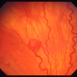
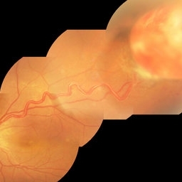
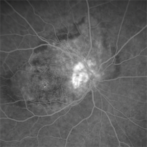
 Optic Nerve/Retinal Capillary Hemangioma
Aug 12 2021 by Stefanie Palmer
Optic Nerve/Retinal Capillary Hemangioma of the Right eye.
Photographer: Stefanie Palmer, CRA
Condition/keywords: hemangioma, optic nerve
-
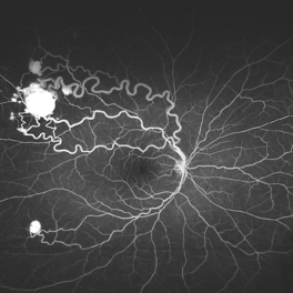
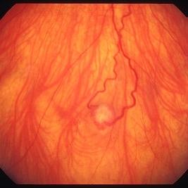
 Retinal capillary hemangiomas 3
Jan 11 2013 by Alex P. Hunyor, MD
Retinal capillary haemangiomas, left superior periphery, in a 20 year old female with von Hippel-Lindau disease.
Condition/keywords: hemangioma, Von Hippel-Lindau
-
 Von Hippel Lindau with retinal capillary hemangioma
Nov 2 2023 by Marcelo Zas, MD PhD
30-year-old female patient diagnosed with Syndrome VHL (Von Hippel Lindau). Stage II. In the first wide-field retinography of the right eye we can observe the exophytic retinal hemangiomas, rounded, slightly delimited, located in the peripheral retina in the upper and lower temporal quadrants and due to the exudation produced by them, hard exudates are observed in the star hemisphere, affecting the macula.
Photographer: Mariano Cotic MD
Imaging device: Silverstone SS OCT Optos
Condition/keywords: abnormal retinal vessel
-
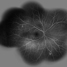
 Von Hippel Lindau With Retinal Capillary Hemangioma
Nov 2 2023 by Marcelo Zas, MD PhD
This wide-field AGF image shows the vascular tumors with their corresponding afferent and efferent vessels.
Photographer: Mariano Cotic MD
Imaging device: Silverstone SS OCT Optos
Condition/keywords: tumor
-
 Von Hippel-Lindau
Sep 1 2014 by Hamid Ahmadieh, MD
Two small retinal capillary hemangiomas detected by wide field FA in the symptom-free right eye of 30-year-old woman with Von Hippel-Lindau.
Photographer: Solmaz Shahmohammad, Negah Eye Center, Tehran, Iran
Condition/keywords: Von Hippel-Lindau
-
 Retinal Capillary Hemangioma
Jan 20 2021 by Jamin S. Brown, MD
Retinal capillary hemangioma, OD.
Photographer: Stefanie Palmer CRA, Retina Vitreous Surgeons of CNY
Condition/keywords: fluorescein angiogram (FA)
-
 Retinal Capillary Hemangioma
Dec 12 2019 by David L Kilpatrick, MD
This is a wide-field color fundus photo showing two distinct retinal capillary hemangiomas. A visually significant epiretinal membrane is also present. Work up with gene testing was negative for VHL. The plan is to proceed with PDT of the two separate lesions (half fluence for the peripapillary lesion), followed by cryotherapy / photocoagulation.
-
 Retinal Capillary Hemangioma
Feb 10 2016 by Claudia G Hooten, MD
Fundus photo of 47-year-old male with PVR retinal detachment 2 months post cryotherapy of RCH.
Photographer: Mark D. Clark
-
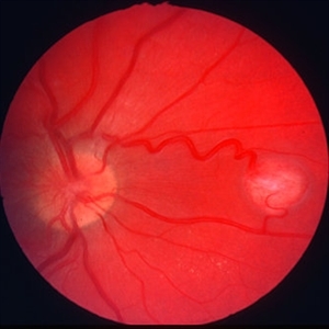
 Retinal Capillary Hemangioma
Mar 21 2013 by Yusuke Oshima, MD, PhD
A peripheral retinal capillary hemangioma.
Photographer: Yusuke Takada, Osaka University Graduate School of Medicine
-
 Retinal capillary hemangioma 2
Jan 11 2013 by Alex P. Hunyor, MD
Retinal capillary haemangioma, right inferior periphery, in a 20-year-old female with von Hippel-Lindau disease.
Condition/keywords: hemangioma, Von Hippel-Lindau
-
 Atypical Arterial Beading Secondary to Retinal Capillary Hemangioma
Jan 28 2020 by Sophia El Hamichi, MD
Image 1: Fundus picture montage depicting RCH with feeder and drainer vessels. Note the unusual beaded appearance of the arterioles. Image 2: 2A: Arterial phase of fluorescein angiography shows early filling of the arteriole. 2B: Arterio-venous phase highlighting the sausage appearance of the arterioles beading.
Condition/keywords: arterial beading, fluorescein angiogram (FA), retinal capillary hemangioblastoma
-
 Capillary Hemangioma
Mar 27 2019 by Gary R. Cook, MD, FACS
White male with a retinal capillary hemangioma OD secondary to neurofibromatosis.
Condition/keywords: neurofibromatosis, retinal hemangioma
-
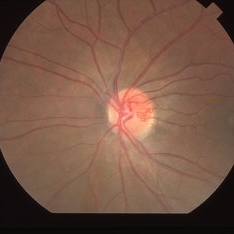 Capillary Hemangioma
Mar 27 2019 by Gary R. Cook, MD, FACS
28-year-old asymptomatic white female with a retinal capillary hemangioma of the optic disc OS; V.A.= 20/20-1.
Imaging device: Topcon VT-50
Condition/keywords: optic disc
-
 Capillary Hemongima, Coat's Response
May 2 2013 by Henry J. Kaplan, MD
Coat's response as exudation in the macula in the same patient with retinal capillary hemangioma. Notice the dilated feeder vessles from the optic nerve infriorly; #2.
Condition/keywords: Coats' disease, Von Hippel-Lindau
-
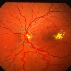
 Hemangioma Retina
Apr 15 2023 by Lulwa El Zein, MD
40 year old female with a incidental finding of retinal capillary hemangioma.
Photographer: Lulwa El Zei, Mayo clinic, Rochester MN
Condition/keywords: fluorescein angiogram (FA), hemangioma, retina
-
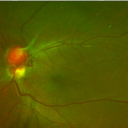
 Juxtapapillary Retinal Capillary Hemangioblastoma
Oct 16 2025 by Sara Mayoral Sánchez
A hyperfluorescent lesion located superotemporal to the optic disc, consistent with a juxtapapillary retinal capillary hemangioma.
Photographer: Sara Mayoral Sánchez, H.U.Puerta del Mar, Cádiz
Condition/keywords: angiography with fluorescein, hemangioma, optic disc, retina capillary hemangioblastoma
-
 RCH-OD
Jul 28 2023 by Mohammadkarim Johari
15 year old girl with bilateral vision loss, in right eye a nodular, orange-colored lesions that grow in the outer layers of the retina is seen in supra-temporal quadrant of retina. Retinal capillary hemangioma is a benign retinal hamartoma that may be associated with von Hippel-Lindau (VHL) disease
Photographer: Mohammadkarim Johari, Shiraz university of medical science
Condition/keywords: retinal capillary hemangioblastoma
-
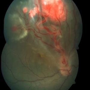 Retinal Capillary Hemangioblastoma
Oct 6 2015 by Pukhraj P Rishi, MBBS, MS, DO, FRCS, FRCSEd, FASRS, FACS
Fundus photograph of an 18-year-old Asian Indian male with multiple retinal capillary hemangiomas with sub retinal fluid.
Photographer: M S KRISHNA
Imaging device: Zeiss FF4
Condition/keywords: retinal angioma, tumor, Von Hippel-Lindau
-
 Retinal capillary hemangioma
Jan 11 2013 by Alex P. Hunyor, MD
Retinal capillary haemangioma nasal to optic disc, right eye.
Condition/keywords: Von Hippel-Lindau
-
---thumb.jpg/image-square;max$300,300.ImageHandler) Retinal Capillary Hemangioma
Retinal Capillary Hemangioma
May 3 2013 by Jerald A. Bovino, MD
Fundus photo of a retinal capillary hemangioma after treatment.
-
---thumb.jpg/image-square;max$300,300.ImageHandler) Retinal Capillary Hemangioma
Retinal Capillary Hemangioma
May 3 2013 by Jerald A. Bovino, MD
Fundus photo of a retinal capillary hemangioma after treatment of the feeder vessel.
Condition/keywords: feeder vessel
-
 Retinal Capillary Hemangioma
May 3 2013 by Jerald A. Bovino, MD
Fundus photo of a retinal capillary hemangioma after treatment of the feeder vessel.
Condition/keywords: feeder vessel
-
 Retinal Capillary Hemangioma
May 31 2017 by S. Natarajan, MD, FASRS, FRCS (GLASGOW) , FICO, D.Sc, FELA
Fundus photograph of an 21-year-old male with double angioma before undergoing laser photo ablation.
Photographer: Ms. Ashwini Borde
Imaging device: Carl Zeiss 450 Plus IR
Condition/keywords: angioma, disc
-
 Retinal Capillary Hemangioma
Sep 9 2021 by Jesus Lozano, MD
60 year-old woman with a Peripheral RCH treated with laser photocoagulation.
Photographer: Yair Bet Yosef, Hadassah Medical Center. Israel
Imaging device: Optos Silverstone
Condition/keywords: abnormal retinal vessel, anomalous vessels, dilated tortuous vessels, hemangioma, retina
-
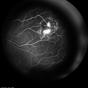 Retinal Capillary Hemangioma
Feb 18 2016 by Hashim Ali Khan, OD, FAAO
FA of 23-year-old-woman with RCH as part of spectrum of Von Hippel- Lindau
Imaging device: Heidelberg Spectralis
Condition/keywords: Von Hippel-Lindau
 Loading…