Search results (33 results)
-
 Myopic retinopathy
Dec 27 2021 by Eduardo Javier Pinuer Alvarado
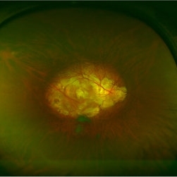
Fundus photograph of an 50-year-old man with myopic retinopathy, posterior staphyloma, myopic chorioretinal atrophy and tilted and oblique disc.
Photographer: Eduardo Pinuer A, Universidad Austral de Chile.
Imaging device: CR-2 AF Digital Non-Mydriatic Retinal Camera, Canon.
Condition/keywords: myopic chorioretinal atropthy, myopic retinopathy, posterior staphyloma, retinopathy
-
 Posterior Staphyloma
Aug 3 2017 by Eitae Kim, MD
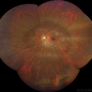
UWF fundus photograph of 35-year-old male with unilateral posterior staphyloma.
Photographer: Eitae Kim, BOIM retina center, Pureun eye hospital
Condition/keywords: ultra-wide field imaging
-
 Myopic Degeneration
Jul 3 2018 by Armando L. Oliver, MD
Myopic Degeneration
Photographer: Moises Castro
Imaging device: Optos California
Condition/keywords: pathologic myopia, posterior staphyloma
-
 Dislocated Intraocular Lens, Tilted Disc, Posterior Staphyloma.
Aug 18 2021 by Jesus Lozano, MD
Fundus photograph of 80-year-old woman, single eye with left eye posterior dislocation of lens, tilted disc and posterior staphyloma.
Photographer: Yair Bet Yosef, Hadassah Medical Center. Israel
Imaging device: Optos Silverstone
Condition/keywords: posterior dislocation of lens, posterior staphyloma, tilted disc
-
 High Myopia
Jun 14 2018 by Mitzy E Torres Soriano, MD
Fundus photograph (left eye) of a female patient with high myopia, chorioretinal atrophy, pigmentary changes and posterior staphyloma.
Photographer: Mitzy Torres Soriano
Condition/keywords: chorioretinal atrophy, high myopia, posterior staphyloma
-
 High Myopia with Posterior staphyloma
Nov 7 2023 by Harsh Vardhan Singh, MS
27-year old with both eyes high myopia & posterior staphyloma with left eye peripheral lattice degeneration & white without pressure
Photographer: Harsh Vardhan Singh
Imaging device: Clarus 700
Condition/keywords: lattice degeneration, myopia, peripheral lattice degeneration, posterior staphylomaloma, white without pressure
-
 Macular Hole Retinal Detachment Over a Posterior Staphyloma
Dec 31 2016 by Linda A Cernichiaro- Espinosa, MD
Macular hole retinal detachment over a posterior staphyloma of pathologic myopia.
Photographer: Linda A Cernichiaro
Imaging device: Optos
Condition/keywords: degenerative myopia, high myopia, macular hole, myopic eye, posterior staphyloma, vitreoretinal degeneration
-
 Myelinated Nerve Fibers and Possibly Posterior Staphyloma
Feb 19 2013 by From the Collections of Thomas M. Aaberg, MD and Thomas M. Aaberg Jr., MD
Findings are bilateral.
Condition/keywords: color photo, myelinated nerve fibers
-
 Myelinated Nerve Fibers and Possibly Posterior Staphyloma
Feb 19 2013 by From the Collections of Thomas M. Aaberg, MD and Thomas M. Aaberg Jr., MD
Findings are bilateral.
Condition/keywords: color photo, myelinated nerve fibers
-
 Myelinated Nerve Fibers and Possibly Posterior Staphyloma
Feb 19 2013 by From the Collections of Thomas M. Aaberg, MD and Thomas M. Aaberg Jr., MD
Findings are bilateral.
Condition/keywords: myelinated nerve fibers
-
 Myelinated Nerve Fibers and Possibly Posterior Staphyloma
Feb 19 2013 by From the Collections of Thomas M. Aaberg, MD and Thomas M. Aaberg Jr., MD
Findings are bilateral.
Condition/keywords: color photo, myelinated nerve fibers
-
 Myelinated Nerve Fibers and Possibly Posterior Staphyloma
Feb 19 2013 by From the Collections of Thomas M. Aaberg, MD and Thomas M. Aaberg Jr., MD
Findings are bilateral.
Condition/keywords: color photo, myelinated nerve fibers
-
 Myopic Degeneration
Jul 3 2018 by Armando L. Oliver, MD
Myopic Degeneration
Photographer: Moises Castro
Imaging device: Optos California
Condition/keywords: pathologic myopia, posterior staphyloma
-
 Myopic Degeneration
Jul 3 2018 by Armando L. Oliver, MD
FAF
Photographer: Moises Castro
Imaging device: Optos California
Condition/keywords: pathologic myopia, posterior staphyloma
-
 Myopic Degeneration
Jul 3 2018 by Armando L. Oliver, MD
FAF
Photographer: Moises Castro
Imaging device: Optos California
Condition/keywords: pathologic myopia, posterior staphyloma
-
 Myopic Degeneration
Jul 3 2018 by Armando L. Oliver, MD
Late Views IVFA
Photographer: Moises Castro
Imaging device: Optos California
Condition/keywords: pathologic myopia, posterior staphyloma
-
 Myopic Degeneration
Jul 3 2018 by Armando L. Oliver, MD
Late views IVFA.
Photographer: Moises Castro
Imaging device: Optos California
Condition/keywords: pathologic myopia, posterior staphyloma
-
 Myopic Degeneration
Oct 4 2014 by Mehul A Shah
A 40-year-old male presented with complaint of gradual diminished vision.
Photographer: Drashti Netralaya,Dahod
Imaging device: Zeiss ff450
Condition/keywords: posterior staphyloma
-
 Myopic Degeneration
Dec 9 2024 by Virginia Gebhart
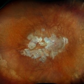
67 year old female with myopic degeneration. Posterior staphylomas are stable. VA limited by extensive chorioretinal atrophy. BCVA 20/160 (ecc)
Photographer: Virginia Gebhart, Retina Consultants of Carolina
Imaging device: Optos California
Condition/keywords: chorioretinal atrophy, myopic degeneration, staphyloma
-
 Myopic Traction Maculopathy
May 31 2014 by Rameez N Hussain, MD
Color photograph of macular detachment in a posterior staphyloma - myopic traction maculopathy (MTM).
Photographer: Rameez N Hussain MD, Vitreo Retinal Services, Giridhar Eye Institute, Cochin, India
Imaging device: Zeiss
Condition/keywords: high myopia, macular detachment, myopic traction maculopathy, pathologic myopia, posterior staphyloma
-
 Myopic Traction Maculopathy
May 31 2014 by Rameez N Hussain, MD
Spectral domain optical coherence tomography of macular detachment in posterior staphyloma - myopic traction maculopathy (MTM).
Photographer: Rameez N Hussain MD, Vitreo Retinal Services, Giridhar Eye Institute, Cochin, India
Imaging device: Heidelberg Spectralis
Condition/keywords: high myopia, macular detachment, myopic traction maculopathy, pathologic myopia, posterior staphyloma
-
 Partial Vitreous Separation in a High Myope With a Posterior Staphyloma
Dec 10 2012 by Yale L. Fisher, MD
This B-scan demonstrates a partial PVD. A posterior vitreous detachment (PVD) may occur in a normal aging eye or may be associated with pathology such as vitreous hemorrhage or inflammation. In a normal eye, as in this example, the PVD appears as a thin and smooth line (arrow) on B-scan. When the globe is moved voluntarily by the patient, real time echography demonstrates a quick jerky motion of the sheet-like echo with movements continuing after the globe movement has ceased. This is helpful in differentiating a PVD from a retinal detachment, which typically has a slower undulating pattern of motion. If there was presence of blood or inflammatory debris associated with the PVD, the echogenic line might appear thicker, especially in the most gravity dependent portions of the globe (i.e., posterior and inferior).
Condition/keywords: video
-
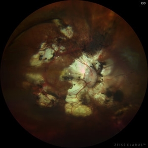 Pathological Myopia with posterior pole retinal detachment
Jul 31 2023 by Harsh Vardhan Singh, MS
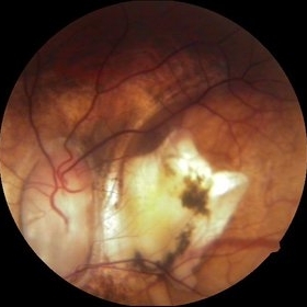
45-year female with right eye re-detachment with pathological myopia & posterior pole RRD with open break
Photographer: Dr Harsh Vardhan Singh, AIIMS, Guwahati
Imaging device: Zeiss clarus 700
Condition/keywords: pathologic myopia, posterior pole lesion, posterior staphyloma, rrd
-
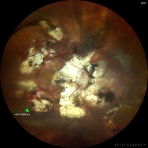 Pathological Myopia with posterior pole retinal detachment & new open break
Jul 31 2023 by Harsh Vardhan Singh, MS
45-year female with redetachment & new break
Photographer: Dr Harsh Vardhan Singh, AIIMS, Guwahati
Imaging device: Zeiss Clarus 700
Condition/keywords: pathologic myopia, posterior staphyloma, retinal break, rrd
-
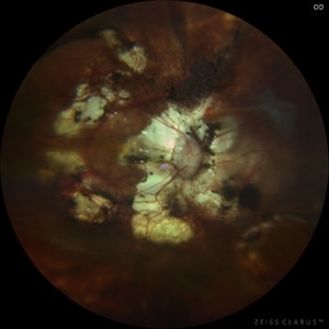 Pathological Myopia with posterior pole retinal detachment & new open break
Jul 31 2023 by Harsh Vardhan Singh, MS
45-year female with redetachment & new break
Photographer: Dr Harsh Vardhan Singh, AIIMS, Guwahati
Imaging device: Zeiss Clarus 700
Condition/keywords: pathologic myopia, posterior staphyloma, retinal break, rrd
 Loading…