Search results (56 results)
-
 Iris Pigmented Lesion
Apr 27 2018 by Mark Lazcano
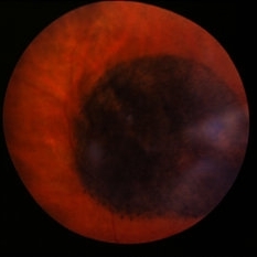
Gonio photograph of 20-year-old male with pigmented iris lesion consistent with melanocytoma
Photographer: mark Lazcano,University of Miami , Bascom Palmer Eye Institute
Imaging device: gonio Prism
Condition/keywords: pigmented lesion
-
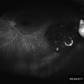
 Adenocarcinoma Arising from CHRPE
Sep 17 2015 by Marc C. Peden, MD
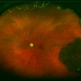
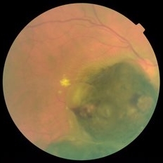
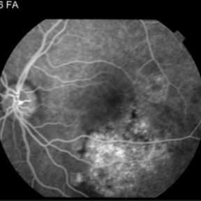
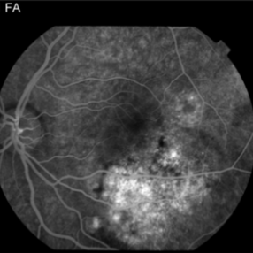
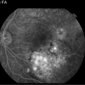
49-year-old female referred for presumed ocular melanoma. On examination was noted to have darkly pigmented lesion in the temporal retina of left eye. Lesion had characteristic scalloped edges with central lacunae, however, on ultrasonography was noted to have 1.8mm of elevation with high internal reflectivity. IVFA shows absence of dual circulation with areas of window defect. Findings were consistent with those described by Shields et al., in their April 2001 article in Archives of Ophthalmology.
Photographer: Janet Traynom
Imaging device: Optos P200MA
Condition/keywords: adenocarcinoma arising from CHRPE
-
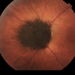
 Melanocytoma of the Optic Nerve
Apr 6 2024 by Hector Gabriel Moreno Solano, MD, MHA
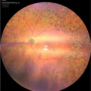
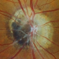
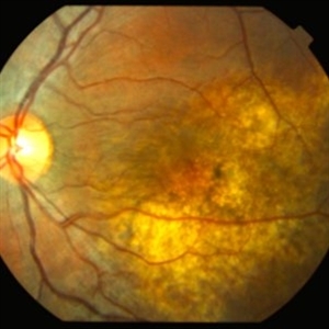
Fundus photograph of a 57-year-old male presented for an ophthalmological evaluation with a chief complaint of progressive visual loss. Indirect ophthalmoscopy revealed proliferative diabetic retinopathy, without macular edema, and a hyperpigmented lesion at the optic disc which corresponds to a melanocytoma.
Photographer: Héctor Gabriel Moreno-Solano
Imaging device: Clarus 700
Condition/keywords: diabetic retinopathy, intraocular tumor, melanocytoma, optic nerve
-
 Melanocytoma of the Optic Nerve
Apr 6 2024 by Hector Gabriel Moreno Solano, MD, MHA
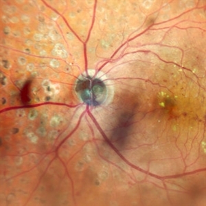
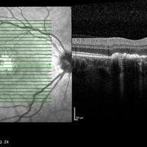
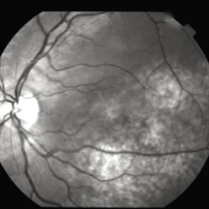
Optic Nerve laser scan image reconstruction of a 57-year-old male presented for an ophthalmological evaluation with a chief complaint of progressive visual loss. Indirect ophthalmoscopy revealed proliferative diabetic retinopathy, without macular edema, and a hyperpigmented lesion at the optic disc which corresponds to a melanocytoma.
Photographer: Héctor Gabriel Moreno-Solano, MD, MHA
Imaging device: Mirante
Condition/keywords: intraocular tumor, macular edema, melanocytoma, optic nerve
-
 Sunset Glow Fundus
May 15 2022 by Manuel Ángel Alcántara Delgado, MD
Optomap ultra-widefield retinal imaging of an 35-year-old woman showed sunset glow fundus, multiple nummular chorioretinal atrophic lesions, macular subretinal fibrosis and pigment clumping in chronic recurrent stage of Vogt-Koyanagi-Harada disease.
Photographer: Manuel Ángel Alcántara Delgado. Conde de Valenciana.
Condition/keywords: abnormal retina, benign pigmented lesions, pigment clumps, retinal fibrosis, uveitis, Vogt-Koyanagi-Harada
-
 Optic Disc Melanocytoma
Sep 14 2023 by Ben Serar
Fundus photograph showing hyper pigmented lesion at the optic disc in a case of Optic disc melanocytoma.
Condition/keywords: optic disc melanocytoma
-
 Unilateral Acute Idiopathic Maculopathy OCT Macula
May 7 2019 by William Ensor
A 37-year-old female presented with a two-week history of vision loss in the right eye. She experienced a flu-like illness including rash on the hands, feet, and mouth 2 days prior to her vision change. Her 3-year-old son had a similar illness diagnosed as hand, foot, and mouth disease by his pediatrician one week prior. Her visual acuity was 20/150 of the right eye, and 20/20 of the left eye. On dilated fundus examination, the left eye was unremarkable; the right eye revealed a circular, variably pigmented lesion of the macula. OCT imaging showed areas of RPE loss and clumping, with overlying loss of the photoreceptor layer. Fluorescein angiography showed central and peripheral hyperfluorescence consistent with window defect, and blockage in area of RPE loss. No treatment was initiated at this time. The patient returned 10 days later; her visual acuity improved to 20/50 in the right eye. Dilated fundus exam showed increased pigmentation of the macular lesion. OCT of the right eye showed further RPE clumping without recovery of the photoreceptor layer, despite her improved visual acuity.
Condition/keywords: unilateral acute idiopathic maculopathy
-
 Bear Tracks
Dec 31 2012 by Raj K. Maturi, MD
Photographer: Tom Steele, CRA Midwest Eye Institute Indianapolis, Indiana
Imaging device: Topcon 50ex 50 degree field
Condition/keywords: bear tracks, benign pigmented lesions, congenital hypertrophy of the retinal pigment epithelium (CHRPE), OD
-
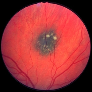
 Unilateral Acute Idiopathic Maculopathy Fundus
May 7 2019 by William Ensor
A 37-year-old female presented with a two-week history of vision loss in the right eye. She experienced a flu-like illness including rash on the hands, feet, and mouth 2 days prior to her vision change. Her 3-year-old son had a similar illness diagnosed as hand, foot, and mouth disease by his pediatrician one week prior. Her visual acuity was 20/150 of the right eye, and 20/20 of the left eye. On dilated fundus examination, the left eye was unremarkable; the right eye revealed a circular, variably pigmented lesion of the macula. OCT imaging showed areas of RPE loss and clumping, with overlying loss of the photoreceptor layer. Fluorescein angiography showed central and peripheral hyperfluorescence consistent with window defect, and blockage in area of RPE loss. No treatment was initiated at this time. The patient returned 10 days later; her visual acuity improved to 20/50 in the right eye. Dilated fundus exam showed increased pigmentation of the macular lesion. OCT of the right eye showed further RPE clumping without recovery of the photoreceptor layer, despite her improved visual acuity.
Condition/keywords: unilateral acute idiopathic maculopathy
-
 Melanocytoma with Choroidal Melanoma
Oct 8 2012 by Susanna S. Park, MD, PhD
Fundus photograph of a 75-year-old woman with a slowly growing pigmented lesion.
Photographer: Ellen Redenbo, University of California Davis Eye Center
Condition/keywords: melanocytoma
-
 61-Year-Old Man With Large Peripheral CHRPE
Dec 9 2017 by Timothy S Fuller, MD
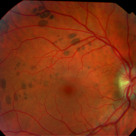
61-year-old man presented for evaluation of pigmented retinal lesion. Found to have a large, peripheral CHRPE with characteristic lacunae, sharp margins, and lack of elevation.
Condition/keywords: benign pigmented lesions, congenital hypertrophy of the retinal pigment epithelium (CHRPE), lacunae
-
 Melanocytoma of Optic Disc
Nov 3 2023 by Virginia Gebhart
69 year-old female with pigmented lesion that covers the optic nerve. Patient has been aware for over 30 years. Remains stable and unchanged
Photographer: Virginia Gebhart
Imaging device: Topcon
Condition/keywords: benign melanocytoma, Melanocytoma, optic disc melanocytoma
-
 Retinal hyperplasia
Feb 19 2018 by JEFFERSON R SOUSA, Tecg.º (Biomedical Systems Technology)
Female patient, 28 years in monitoring to control a hyperpigmented lesion in the temporal retina of the right eye.
Photographer: Photographer JEFFERSON ROCHA DE SOUSA, Clinic Dr. Marco Antonio Albhy Oftalmology, Institute Dr. Suel Abujamra São Paulo-Brazil
Imaging device: Retinografo Topcin TRC-NW6S. Mosaic, Flash 25.
Condition/keywords: hyperplasia, hyperplastic retinal pigment epithelium (RPE)
-
 Choroidal naevus
Jan 11 2013 by Alex P. Hunyor, MD
Choroidal naevus with overlying drusen
Condition/keywords: benign pigmented lesions, choroidal nevus
-
 Adenocarcinoma Arising from CHRPE
Sep 17 2015 by Marc C. Peden, MD
49-year-old female referred for presumed ocular melanoma. On examination was noted to have darkly pigmented lesion in the temporal retina of left eye. Lesion had characteristic scalloped edges with central lacunae, however, on ultrasonography was noted to have 1.8mm of elevation with high internal reflectivity. IVFA shows absence of dual circulation with areas of window defect. Findings were consistent with those described by Shields et al., in their April 2001 article in Archives of Ophthalmology.
Photographer: Janet Traynom COT
Imaging device: Optos P200MA
Condition/keywords: adenocarcinoma arising from CHRPE
-
---thumb.jpg/image-square;max$300,300.ImageHandler) Benign Melanocytoma of the Optic Disc
Benign Melanocytoma of the Optic Disc
Jan 11 2013 by Hyung-Woo Kwak, MD
Fundus photography of optic disc showing a dark pigmented lesion.
Photographer: Dongho Kang, Kyung Hee Univsersity Hospital, Seoul
Imaging device: Zeiss f 450 plus
Condition/keywords: benign melanocytoma
-
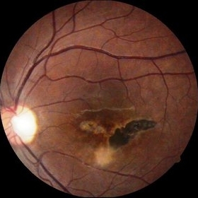 Central Chorioretinitis
Oct 3 2014 by Mehul A Shah
A 30-year-old female presented with complaint of sudden loss of vision OS before 2 months, on examination she was found to have pigmented lesion.
Photographer: Drashti Netralaya,Dahod
Imaging device: Zeiss ff450
-
 Choroidal Melanocytosis
Sep 14 2023 by Ben Serar
Fundus photograph showing hyper pigmented lesion near the disc, at the level of the choroid, in a case of choroidal melanocytosis.
Condition/keywords: choroidal melanocytosis
-
 Choroidal Melanoma
Jan 29 2015 by H. Michael Lambert, MD
Dark pigmented lesion in retina with elevation.
-
 Choroidal Melanoma
May 15 2014 by Mitzy E Torres Soriano, MD
Fundus photograph of a 55-year-old male with pigmented, elevated lesion involving optic nerve. with exudation, hemorrhage and subretinal fluid.
Photographer: Mitzy E Torres Soriano, Centro Medico Cagua, Venezuela
Imaging device: Retinal camera TRC-NW8, TOPCON
Condition/keywords: choroidal nevus, pigmented lesion
-
 Choroidal Osteoma
Nov 21 2014 by Thomas A. Ciulla, MD, MBA, FASRS
This 13-year-old girl presented with mild painless progressive blurring of central vision left eye over the past several months. Visual acuity was 20/25. In the affected left eye, retinal examination revealed a relatively flat, lightly pigmented lesion, with well-defined and scalloped edges. Clumps of associated pigment were noted.
Photographer: Thomas Steele
Condition/keywords: choroidal neovascular membrane (CNVM), choroidal neovascularization (CNV), choroidal osteoma, macular choroidal osteoma
-
 Choroidal Osteoma
Nov 21 2014 by Thomas A. Ciulla, MD, MBA, FASRS
This 13-year-old girl presented with mild painless progressive blurring of central vision left eye over the past several months. Visual acuity was 20/25. In the affected left eye, retinal examination revealed a relatively flat, lightly pigmented lesion, with well-defined and scalloped edges. Clumps of associated pigment were noted. This OCT image shows subretiinal fluid just inferior to the fovea. Choroidal osteoma can be associated with the development of subretinal neovascularization (particularly at the edges of the osteoma).
Photographer: Thomas Steele
Condition/keywords: choroidal neovascular membrane (CNVM), choroidal neovascularization (CNV), choroidal osteoma, macular choroidal osteoma
 Loading…