Search results (5 results)
-
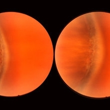 Normal Temporal Ora Serrata
Nov 9 2012 by Norman Byer
This is the normal temporal ora serrata in a 26-year-old man. Note the typical ragged moth-eaten appearance caused by peripheral cystoid degeneration. This appearance may be present in infants but is always present beyond the age of eight years.
Condition/keywords: ora serrata, peripheral cystoid degeneration
-
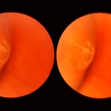 Meridional Fold
Nov 9 2012 by Norman Byer
This is the same lesion as in the previous photograph. With the scleral indentation placed more posterior, we now can see that the fold ends over a small collection of subretinal fluid and that there is a very tiny retinal hole just below the posterior end of the retinal fold.
Condition/keywords: peripheral cystoid degeneration, retinal fold, retinal hole, scleral indentation, subretinal fluid
-
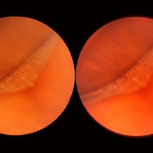 Meridional Fold
Nov 9 2012 by Norman Byer
The next two photographs are of the same lesion in a 28-year-old woman. This view shows a sloping retinal mound with a radial retinal fold in the center. This is not a typical meridional fold for it stops short of the ora serrata and there is no dentate process. The upper temporal ora serrata and pars plana are well shown and peripheral cystoid degeneration is present posterior to the ora.
Condition/keywords: ora serrata, pars plana, peripheral cystoid degeneration, radial retinal fold, sloping retinal mound
-
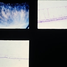
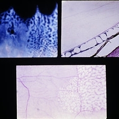 Slide 9-47
Feb 26 2019 by Lancaster Course in Ophthalmology
Typical peripheral cystoid degeneration. A small, round hole within the area of cystoid degeneration is present (upper left). In section the cysts are located mostly in the outer plexiform layer (upper left). A nondigestion flat preparation of the retina shows the cystic spaces, which have coalesced to form meridionally oriented tunnels (lower view).
Condition/keywords: peripheral cystoid degeneration
-
 Slide 9-48
Feb 26 2019 by Lancaster Course in Ophthalmology
Typical peripheral cystoid degeneration (PCD) just posterior to the ora serrata, and reticular peripheral cystoid degeneration located posterior to the typical PCD. Section of reticular peripheral cystoid degeneration showing cystic spaces in the nerve fiber layer (upper right). Lower view shows junction of typical PCD (left} with reticular PCD (right). (Courtesy of Robert Y Foos, M.D. )
Condition/keywords: peripheral cystoid degeneration
 Loading…