Search results (14 results)
-
 Advanced RP
Nov 5 2024 by rahul saradge
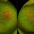
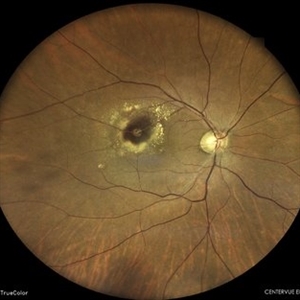
A man, 58, arrived complaining of BOV for both near and distance vision in both eyes, with a 6/9 BCVA in each eye. For a year, the patient had been taking medication for both diabetes and hypertension. In both eyes, the dilated ophthalmoscopic retina revealed waxy disc pallor paired with bony spicules in the mid-periphery. The patient was prescribed spectacles and given counseling regarding the nature of the illness.
Photographer: Lokesh Dukare ,Isha Netralaya Thane
Imaging device: optos
Condition/keywords: bone spicule, optic disc pallor, Optos, Retinitis Pigmentosa
-
---thumb.JPG/image-square;max$300,300.ImageHandler) Anterior Ischaemic Optic Neuropathy
Anterior Ischaemic Optic Neuropathy
Dec 7 2013 by Mallika Goyal, MD
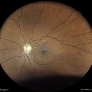
Right eye of a diabetic 65-year-old gentleman with superior half optic disc pallor 2 months following sudden vision drop from anterior ischaemic optic neuropathy. Superior disc pallor corresponds to inferior altitudinal field defect. There was no visual or field improvement following oral steroids.
Photographer: Mallika Goyal, MD, Apollo Health City, Hyderabad, India
Condition/keywords: anterior ischemic optic neuropathy
-
 Behcet's Disease
Nov 25 2012 by Mallika Goyal, MD
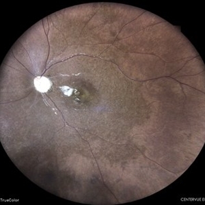
Fundus photograph of right eye of a 23-year-old gentleman with Behcet's Disease shows occlusive retinal vasculitis with optic disc pallor and macular ischaemia. Other eye has similar appearance with no light perception.
Photographer: Mallika Goyal, MD, Apollo Health City, Hyderabad, India
Condition/keywords: occlusive vasculitis
-
---thumb.JPG/image-square;max$300,300.ImageHandler) Behcet's Disease
Behcet's Disease
Nov 25 2012 by Mallika Goyal, MD
Fundus photograph of right eye of a 23-year-old gentleman with Behcet's Disease shows occlusive retinal vasculitis with optic disc pallor and macular ischaemia. Other eye has similar appearance with no light perception.
Photographer: Mallika Goyal, MD, Apollo Health City, Hyderabad, India
Condition/keywords: macular ischemia, occlusive vasculitis
-
 Behcet's Disease
Nov 25 2012 by Mallika Goyal, MD
Fundus photograph of left eye of a 23-year-old gentleman with Behcet's Disease shows occlusive retinal vasculitis with optic disc pallor and macular ischemia. This eye has no light perception; other eye has similar fundus appearance.
Photographer: Mallika Goyal, MD, Apollo Health City, Hyderabad, India
Condition/keywords: macular ischemia, occlusive vasculitis, optic disc pallor
-
 Disc Pallor Status Post NAION
May 7 2024 by Akansha Sharma
Color fundus photograph of a 48 year old male with disc pallor status post NAION.
Photographer: Dr. Akansha Sharma, Bharati Eye Hospital
Condition/keywords: NAION, optic disc pallor
-
 Disc Pallor With Retinal Atrophy Status Post Ischaemic Vascular Event
Apr 8 2024 by Akansha Sharma
Color fundus photograph of a 22 year old female with disc pallor with retinal atrophy status post ischaemic vascular event.
Photographer: Dr. Akansha Sharma, Bharati Eye Hospital
Condition/keywords: inner retinal thinning, optic disc pallor
-
 Dropped Nucleus With Disc Pallor With Posterior Pole Retinal Detachment
Sep 12 2025 by Akansha Sharma
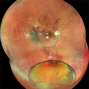
Color fundus photograph of a 60 year old male with a dropped nucleus with disc pallor with posterior pole retinal detachment.
Photographer: DR. AKANSHA SHARMA
Condition/keywords: dropped nucleus, fragmatome, nucleus drop, optic disc pallor, PALE DISC, POSTERIOR POLE RETINAL DETACHMENT, RD, retinal detachment
-
 Hypertensive Retinopathy
Sep 12 2023 by Ben Serar
Fundus photograph of LE showing Disc edema with optic disc pallor, hard exudates with dot-blot haemorrhages at the macula ,along with arteriolar attenuation, in a case of Hypertensive retinopathy.
Condition/keywords: arteriolar attenuation, disc edema, Hard exudates, hypertensive retinopathy
-
 Macular Colobomas in Congenital Zika Syndrome
Sep 26 2020 by Swati Agarwal-Sinha, MD, FASRS
Color fundus picture of the right (OD) and left (OS) eye of 3-day-old female infant with congenital Zika syndrome with bilateral macular colobomatous like chorioretinal atrophy, attenuated vessels, pigmentary changes, and optic disc pallor.
Photographer: Swati Agarwal-Sinha, MD
Condition/keywords: fundus photograph, macular coloboma, zika
-
 Retinal Arterial Macroaneurysm
Jun 5 2024 by Akansha Sharma
Color fundus photograph of a 61 year old hypertensive male with retinal arterial macroaneurysm.
Photographer: Dr. Akansha Sharma, Bharati Eye Hospital
Condition/keywords: optic disc pallor, RAM
-
 Retinitis pigmentosa
Feb 26 2020 by Manuel Ángel Alcántara Delgado, MD
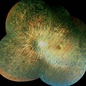
Merged color fundus photograph of a 68-year-old woman with advanced retinitis pigmentosa. It is appreciated bone spicule-shaped pigment deposits, optic disc pallor, retinal atrophy and attenuated retinal vessels.
Photographer: Manuel Ángel Alcántara Delgado
Condition/keywords: choroidal circulation, optic disc pallor, pericentral retinitis pigmentosa, retina, retinitis pigmentosa, retinitis pigmentosa (RP) dystrophy, sector retinitis pigmentosa
-
 Retinitis Pigmentosa
Feb 26 2020 by Manuel Ángel Alcántara Delgado, MD
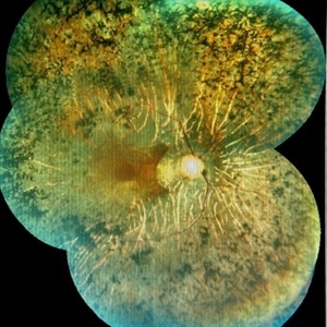
Merged color fundus photograph of a 68-year-old woman with advanced retinitis pigmentosa. It is appreciated bone spicule-shaped pigment deposits, optic disc pallor, retinal atrophy, attenuated retinal vessels and surface wrinkling retinopathy.
Photographer: Manuel Ángel Alcántara Delgado
Condition/keywords: chorioretinal atrophy, choroidal circulation, optic disc pallor, pericentral retinitis pigmentosa, retina, retinitis pigmentosa, retinitis pigmentosa (RP) dystrophy, sector retinitis pigmentosa
-
---thumb.JPG/image-square;max$300,300.ImageHandler) Traumatic Optic Neuropathy
Traumatic Optic Neuropathy
Dec 9 2012 by Mallika Goyal, MD
Right eye of a 23-year-old gentleman 6 months following a road accident. Optic disc pallor with peripapillary chorioretinal scarring suggests traumatic optic neuropathy as the cause of optic atrophy.
Photographer: Mallika Goyal, MD, Apollo Health City, Hyderabad, India
Condition/keywords: traumatic optic neuropathy
 Loading…