Search results (130 results)
-
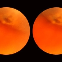
 Puzzle Retinitis
Jan 20 2021 by Jamin S. Brown, MD
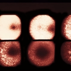
Puzzle artifact after imaging on a smaller field of view with blue light autofluorescence.
Photographer: Stefanie Palmer CRA, Retina Vitreous Surgeons of CNY
Condition/keywords: autofluorescence imaging, normal eye
-
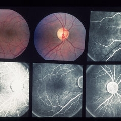
 Normal Eye
Aug 27 2012 by Suber S. Huang, MD, MBA, FASRS
Photographer: Geoffrey Pankhurst, Case Western Reserve University/University Hospitals of Cleveland, Cleveland, OH
Imaging device: Topcon
-
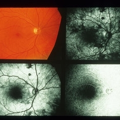
 Normal Eye
Aug 27 2012 by Suber S. Huang, MD, MBA, FASRS
Photographer: Geoffrey Pankhurst, Case Western Reserve University/University Hospitals of Cleveland, Cleveland, OH
Imaging device: Topcon
-
---thumb.jpg/image-square;max$300,300.ImageHandler) Normal Fundus Photo
Normal Fundus Photo
Feb 13 2013 by From the Collections of Thomas M. Aaberg, MD and Thomas M. Aaberg Jr., MD
Normal fundus photo.
Condition/keywords: fundus photograph, normal eye
-
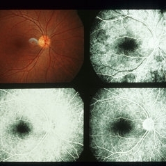
 Normal Eye
Aug 27 2012 by Suber S. Huang, MD, MBA, FASRS
Photographer: Geoffrey Pankhurst, Case Western Reserve University/University Hospitals of Cleveland, Cleveland, OH
Imaging device: Topcon
-
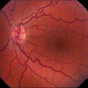 Congénital Venous Tortuosity OS
Mar 13 2013 by Jose Dalma-Weiszhausz, MD
Young patient with routine normal eye exam.
Photographer: José Dalma, MD Dalma & Assoc., Mexico City
Imaging device: Topcon 50VT
Condition/keywords: congenital venous tortuosity
-
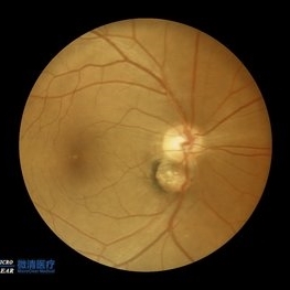 Double disc sign
Oct 13 2022 by Vaibhavi Noticewala, M S Ophthalmology, FVRS
Double disc sign Doubling of the optic disc is rare and can manifest as true or pseudo doubling. Duke-Elder describes duplication of the optic disc as a rare anomaly wherein two discs, each provided with retinal vessels are seen in an otherwise normal eye. Rare cases of true duplication of optic discs with separation of optic nerve into two or more strands have been reported, based either on incidental necropsy findings, demonstration of two optic foramina in the same orbit on x ray, or angioscotomas as indirect evidence of the existence of double optic nerves. Pseudo doubling of the optic discs caused by lesions such as optic disc coloboma, peripapillary chorioretinal coloboma, or inflammatory foci are more common. Our case had Ipsilateral isolated ectatic peripapillary chorioretinal coloboma simulating double optic discs.
Photographer: Priyal Mistry
Condition/keywords: Pseudoduplication of optic disc
-
 Enclosed Ora Bay On The Temporal Side
Nov 9 2012 by Norman Byer
This is another example of an enclosed ora bay on the temporal side. It is surrounded by normal retina and well separated from the ora serrata, which is toward the upper right just beyond the photograph. The yellow nubbin marks an abortive dentate process.
Condition/keywords: abortive dentate process, enclosed ora bay, normal eye, normal retina, ora serrata, temporal retina
-
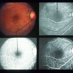
---thumb.jpg/image-square;max$300,300.ImageHandler) Fluoresecein Angiography
Fluoresecein Angiography
Feb 20 2013 by From the Collections of Thomas M. Aaberg, MD and Thomas M. Aaberg Jr., MD
Fluorescein angiography showing a normal venous phase flow shot.
Condition/keywords: normal eye, venous flow phase
-
 Left -Anterior Segment
Aug 10 2020 by RITESH VERMA
Normal anterior segment of the left eye.
Photographer: Dr. Ritesh Verma, Regional institute of Ophthalmology, Rohtak, Haryana, India
Imaging device: CR-2AF CANON
Condition/keywords: anterior segment, normal eye
-
 Looks Pretty Normal
-
 Normal
Jan 7 2015 by H. Michael Lambert, MD
Color photograph of normal fundus.
Condition/keywords: normal eye, normal retina
-
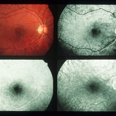
 Normal
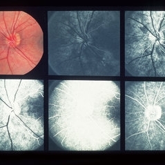
Jul 22 2013 by Howard Schatz, MD
14-year-old white male, C11-Ret Arts.
Condition/keywords: normal eye
-
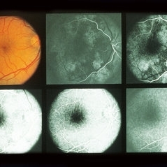
 Normal
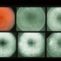
Jul 22 2013 by Howard Schatz, MD
28-year-old white male, right eye 20/15, left eye 20/20.
Condition/keywords: normal eye
-
 Normal
-
 Normal
-
 Normal
-
 Normal
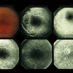
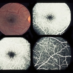
Jul 22 2013 by Howard Schatz, MD
37-year-old black female, 1/10 retinal art 20/200.
Condition/keywords: normal eye
-
 Normal
-
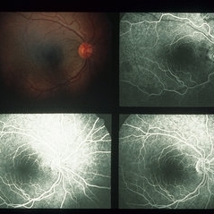
 Normal
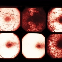
Jul 22 2013 by Howard Schatz, MD
36-year-old white female, right eye 20/40, left eye 20/100.
Condition/keywords: normal eye
-
 Normal
Jul 22 2013 by Howard Schatz, MD
15-year-old white female, right eye 20/40, left eye CF2.
Condition/keywords: normal eye
-
 Normal
-
 Normal
Jul 22 2013 by Howard Schatz, MD
46-year-old, white male, right eye 20/50, left eye 20/50.
Condition/keywords: normal eye
-
 Normal
Jul 22 2013 by Howard Schatz, MD
36-year-old white male, right eye 20/20, left eye 20/32.
Condition/keywords: normal eye
-
 Normal
 Loading…