Search results (60 results)
-
 Cat Scratch
Feb 15 2017 by Hua Gao, MD, PhD, FASRS
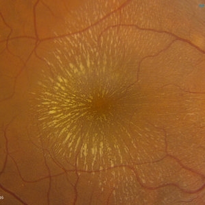
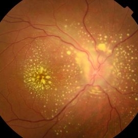
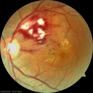
A female patient of 57-year-old presented with neuroretinitis due to cat-scratch disease with positive Bartonella henselae antibodies. Two weeks after symptom onset she developed exudates in a "macular star" pattern.
Condition/keywords: cat scratch retinitis
-
 Central Retinal Vein Occlusion
Jun 21 2025 by Moazzam Parvez
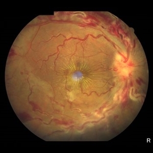
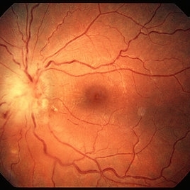
Fundus photograph of a 56 year old male presenting with dilated tortuous vessels with adjoining Hard exudates and macular star.
Photographer: Moazzam Parvez , Netralayam , Kolkata
Imaging device: Topcon Maestro 2
Condition/keywords: CRVO with macular edema, hard exudates, macular star
-
 Macular Star
May 27 2025 by César Adrián Gómez Valdivia, MD
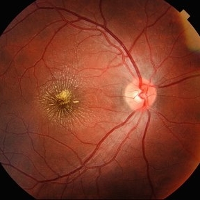
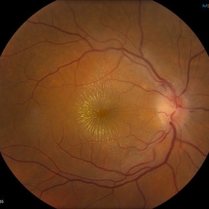
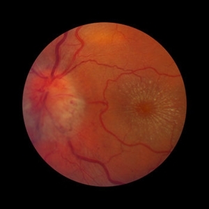
Macular Star found in a 31 year-old male patient with suspected Cat Scratch Disease. Typical intraocular presentations include neuroretinitis with optic nerve edema, macular star formation, and discrete white retinal or choroidal lesions. Findings were unilateral.
Photographer: @eyemissu2
Imaging device: TOPCON TRC-50DX
Condition/keywords: macular star
-
 Infectious Neuroretinitis
May 26 2025 by César Adrián Gómez Valdivia, MD
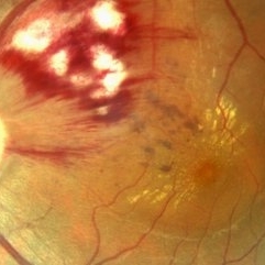
Neuroretinitis found in a 38 year-oldmale patient with IV drugs abuse history. Findings were bilateral. The lipid-rich component of the exudate is able to penetrate into the outer plexiform layer, creating what is clinically seen as a macular star pattern.
Photographer: @eyemissu2
Imaging device: TOPCON TRC-50DX
Condition/keywords: neuroretinitis
-
 Macular Star
Sep 8 2024 by Cesar Augusto Rocha Rojas, MD
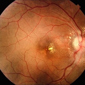
Fundus photograph of a 27-year-old male with hypertensive emergency secondary to chronic kidney disease.
Photographer: Cesar Augusto Rocha Rojas, Hospital General de Zona #20, Instituto Mexicano del Seguro Social (IMSS)
Imaging device: Smartphone, Pan Retinal 2.2 Lens
Condition/keywords: macular star
-
 Optic Disc Edema With Macular Star
Jun 22 2013 by James A Eadie, MD
Fundus photograph montage of a 14-year-old girl with optic disc edema with macular star. Her laboratory work-up was negative for known causes. She improved from 20/200 to 20/40 with observation/an empirical course of doxycycline.
Photographer: Wendy Malmberg-Lorentz
Condition/keywords: neuroretinitis, optic disc edema
-
 Uveitis Posterior
Jul 19 2019 by JEFFERSON R SOUSA, Tecg.º (Biomedical Systems Technology)
A 23-year-old male patient attended the clinic with low vision of the right eye. In the evaluation it presented important fundoscopical alterations like retinal exudations in the posterior pole and nasal retina, aspects of macular star. It was proven that it was a posterior uveitis.
Photographer: JEFFERSON R SOUSA - Study Center and Ophthalmological Research Dr. Andre M V Gomes, Institute Dr. Suel Abujamra São Paulo-Brazil
Imaging device: Topcon TRC-50 DX, Imaginet 4.0, angle de 50 graus. Flash 50w-s
Condition/keywords: uveitis
-
 Toxoplasma Neuroretinitis (Jensen`s Disease)
Feb 25 2013 by Henry J. Kaplan, MD
Toxoplasma neuroretinitis in the left eye of a patient with macular star formation, retinitis adjacent to the optic nerve head with disc swelling.
Condition/keywords: Jensen disease, ocular toxoplasmosis, toxoplasmosis
-
---thumb.jpg/image-square;max$300,300.ImageHandler) Possible CMV Retinitis with Frosted Branch Angiitis
Possible CMV Retinitis with Frosted Branch Angiitis
Feb 14 2013 by From the Collections of Thomas M. Aaberg, MD and Thomas M. Aaberg Jr., MD
Possible CMV Retinitis with frosted branch angiitis appearance and disc edema---late macular star appearance, but diagnosis is not certain.
Condition/keywords: frosted branch angiitis, late macular star, optic disc edema
-
 Bilateral Macular Star
Mar 27 2014 by Jason S. Calhoun
Young female patient in with blurred vision in both eyes. VA is 20/40 in both eyes. Fundus photos show visible macular star centrally in both eyes. This is a result of Bilateral Neuroretinitis due to cat scratch.
Photographer: Jason S. Calhoun, Mayo Clinic Jacksonville, Department of Ophthalmology
Imaging device: TOPCON TRC 50-EX
Condition/keywords: macular star, neuroretinitis
-
 Bilateral Macular Star
Mar 27 2014 by Jason S. Calhoun
Young female patient in with blurred vision in both eyes. VA is 20/40 in both eyes. Fundus photos show visible macular star centrally in both eyes. This is a result of bilateral neuroretinitis due to cat scratch.
Photographer: Jason S. Calhoun, Mayo Clinic Jacksonville, Department of Ophthalmology
Imaging device: TOPCON TRC 50-EX
Condition/keywords: macular star, neuroretinitis
-
 Cat Scratch
Feb 15 2017 by Hua Gao, MD, PhD, FASRS
A female patient of 57-year-old presented with neuroretinitis due to cat-scratch disease with positive Bartonella henselae antibodies. Two weeks after symptom onset she developed exudates in a "macular star" pattern.
Condition/keywords: cat scratch retinitis
-
 HTN / Macular Star / CWS
Feb 17 2015 by David Callanan, MD
HTN / Macular star / CWS.
Condition/keywords: cotton wool spots, hypertension, macular star
-
 HTN / Macular Star / CWS
Feb 17 2015 by David Callanan, MD
HTN / Macular star / CWS.
Condition/keywords: cotton wool spots, hypertension, macular star
-
 Hypertensive Retinopathy
Feb 24 2020 by Brian K. Horsman, MD, FRCS(C) FASRS
Macular star, disc edema, intra vitreous bubble of Bevacizumab
Condition/keywords: bevacizumab, hypertension
-
 Hypertensive Retinopathy
Feb 24 2020 by Brian K. Horsman, MD, FRCS(C) FASRS
Macular star, disc edema, intra vitreous bubble of Bevacizumab
Condition/keywords: bevacizumab, hypertension
-
---thumb.JPG/image-square;max$300,300.ImageHandler) Hypertensive Retinopathy
Hypertensive Retinopathy
Dec 12 2012 by Mallika Goyal, MD
Right eye of a 35-year-old hypertensive gentleman with BP 240/120 mm Hg when first detected a week prior to presentation. Retinal haemorrhages, exudates, macular star and disc edema suggestive of hypertensive retinopathy grade 4.
Photographer: Mallika Goyal, MD, Apollo Hospitals, Hyderabad, India
Condition/keywords: hypertensive retinopathy
-
 Hypertensive Retinopathy
Dec 12 2012 by Mallika Goyal, MD
Left eye of a 35-year-old hypertensive gentleman with BP 240/120 mm Hg when first detected a week prior to presentation. Retinal haemorrhages, exudates, macular star and disc edema suggestive of hypertensive retinopathy grade 4.
Photographer: Mallika Goyal, MD, Apollo Hospitals, Hyderabad, India
Condition/keywords: hypertensive retinopathy
-
 Hypertensive Retinopathy
May 1 2024 by Marco Antonio Sauza
36 year old male with uncontrolled systemic hypertension.
Photographer: MARCO SAUZA CASTILLEJOS
Imaging device: VISUCAM ZEISS
Condition/keywords: choroidal infarction, hypertensive retinopathy, macular star
-
 Hypertensive Retinopathy Grade IV
May 1 2024 by Marco Antonio Sauza
Fundus photograph of an 36-year-old male with uncontrolled systemic hypertension, >200/100mmhg, presenting decreased vision in the left eye.
Photographer: MARCO SAUZA CASTILLEJOS
Imaging device: VISUCAM ZEISS
Condition/keywords: choroidal infarction, hypertensive retinopathy, macular star
-
 LE WIDEFIELD FUNDUS PHOTOGRAPH OF HYPERTENSIVE RETINOPATHY GRADE IV
Sep 27 2023 by ANKIT JAIN
WIDEFIELD IMAGE OF RE OF 14 YEARS OLD MALE RECENTLY DIAGNOSED WITH MALIGNANT HYPERTENSION SHOWING MACULAR STAR, DISC EDEMA IN A CASE OF HYPERTENSIVE RETINOPATHY GRADE IV.
Photographer: DR ANKIT JAIN
Imaging device: MIRANTE
Condition/keywords: Disc Edema, hypertensive retinopathy, macular star
-
 Leptospirosis Neuroretinitis
May 30 2014 by Mitzy E Torres Soriano, MD
50-year-old man, presented with sudden onset of reduced vision in the left eye. Visual acuity (VA) was count fingers. Fundoscopic examination revealed soft exudation adjacent the optic nerve, macular edema with hard exudates in star shape arrangement and retinal vasculitis. OCT confirmed macular edema. There were no systemic symptoms. History of alcoholism and crack cocaine addiction. Systemic work up revealed a positive leptospira. He was treated with oral doxycicline (100mg twice daily) and prednisona (1mg/kg with gradual taper) for two weeks. Follow up at six months showed an improvement of VA to 20/60 with partial resolution of clinical findings at fundoscopic exam. Leptospirosis should be ruled out in every case of neuroretinitis.
Photographer: Mitzy E. Torres Soriano, MD; Centro medico Cagua-Estado Aragua. Venezuela
Imaging device: Retinal Camera TRC-NW8, TOPCON
Condition/keywords: leptospirosis, macular star, neuroretinitis, retinal vasculitis
-
 Macular Star
Jul 14 2013 by Jason S. Calhoun
Macular star with no optic nerve edema.
Photographer: Jason S. Calhoun, Department of Ophthalmology, Mayo Clinic Jacksonville, Florida
Imaging device: TOPCON TRC 50-EX
-
---thumb.jpg/image-square;max$300,300.ImageHandler) Macular Star
Macular Star
Feb 13 2013 by From the Collections of Thomas M. Aaberg, MD and Thomas M. Aaberg Jr., MD
Patient AR.
-
 Macular Star
Oct 19 2016 by Chuck Terranova
Fundus photo of macular star.
Photographer: Chuck Terranova, The Ross Eye Institute, Buffalo, NY.
Condition/keywords: fundus photograph, macular star, retina
 Loading…