Search results (43 results)
-
 Macular Coloboma OS
Sep 26 2012 by Jose Dalma-Weiszhausz, MD
25-year-old male with poor vision since birth OU.
Photographer: José Dalma, MD, Dalma & Asoc. Mexico City, Mexico
Condition/keywords: macular coloboma
-
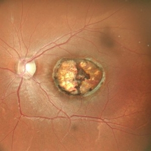
 MACULAR COLOBOMA
Oct 15 2022 by Akansha Sharma
COLOUR FUNDUS PHOTOGRAPH OF A 32 YEAR OLD MALE WITH MACULAR COLOBOMA
Photographer: Dr. Akansha Sharma-Retina Foundation, Ahmedabad
Condition/keywords: coloboma of macula
-
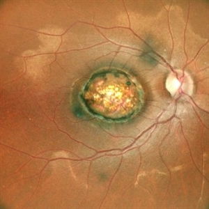
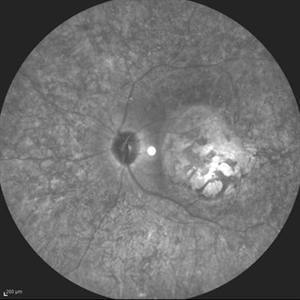
 Macular Coloboma
Jul 17 2024 by Anubhav Chauhan
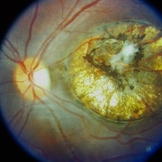
This is fundus photograph of a 30 year male depicting a Macular coloboma in the right eye. The patient had a sharply defined large, yellowish white, coarsely pigmented, atrophic, round crater like defect at the macula. Spectral domain optical coherence tomography confirmed our diagnosis. The serology testing such as serum IgM, IgG for toxoplasma and cytomegalovirus was negative. His systemic examination was normal.
Photographer: Dr Anubhav Chauhan, Department of Ophthalmology, Shri Lal Bahadur Shastri Government Medical College, Nerchowk, District Mandi, Himachal Pradesh, India
Imaging device: Zeiss
Condition/keywords: macula, rare
-
 Macular Coloboma OD
Sep 26 2012 by Jose Dalma-Weiszhausz, MD
25-year-old male with poor vision since birth OU.
Photographer: José Dalma, MD, Dalma & Asoc. Mexico City, Mexico
Condition/keywords: macular coloboma
-
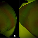 Bilateral Macular Colobomata With Temporal Dragging of Optic Disc
Apr 24 2020 by Dinesh Rungta, MBBS, DNB
Optos ultra-widefield retinal image of a 7-year-old male child showing bilateral macular colobomata with temporal dragging of optic disc.
Photographer: Dr Shivam Madan, Giridhar Eye Institute, Kerala, India
Imaging device: Optos UWF Daytona Plus
Condition/keywords: bilateral macular colobomata, temporal dragging of optic disc
-
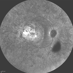 Leber's Congenital Amaurosis
Feb 25 2017 by Hamid Ahmadieh, MD
Infrared image of the right eye of a 25-year-old woman with bilateral macular colobomata and pigmentary retinopathy similar to Leber's congenital amaurosis.
Photographer: Shabnam Poureh, Negah Eye Center, Tehran, Iran
Condition/keywords: bilateral pigmentary retinopathy, infrared image, macular coloboma
-
 Macular Coloboma
Jul 30 2014 by Mallika Goyal, MD
Right fundus of a 20-year-old lady shows macular and the lower part of an extramacular coloboma. VA is stable since early childhood and is 20/200.
Photographer: Mallika Goyal, MD, Apollo Health City, Jubilee Hills, Hyderabad-500033
Condition/keywords: macular coloboma
-
 Macular Coloboma
Jul 30 2014 by Mallika Goyal, MD
Right fundus of a 20-year-old lady shows macular coloboma. VA is stable since early childhood and is 20/200.
Photographer: Mallika Goyal, MD, Apollo Health City, Jubilee Hills, Hyderabad-500033
Condition/keywords: macular coloboma
-
 Macular Coloboma
Jul 30 2014 by Mallika Goyal, MD
Left fundus of a 20-year-old lady shows macular coloboma. VA is stable since early childhood and is 20/25.
Photographer: Mallika Goyal, MD, Apollo Health City, Jubilee Hills, Hyderabad-500033
Condition/keywords: macular coloboma
-
 Macular Coloboma
Jul 30 2014 by Mallika Goyal, MD
Left fundus of a 20-year-old lady shows macular coloboma. VA is stable since early childhood and is 20/25.
Photographer: Mallika Goyal, MD, Apollo Health City, Jubilee Hills, Hyderabad-500033
Condition/keywords: macular coloboma
-
 MACULAR COLOBOMA
Oct 15 2022 by Akansha Sharma
COLOUR FUNDUS PHOTOGRAPH OF A 32 YEAR OLD MALE WITH MACULAR COLOBOMA
Photographer: Dr. Akansha Sharma-Retina Foundation, Ahmedabad
Condition/keywords: coloboma of macula
-
---thumb.jpg/image-square;max$300,300.ImageHandler) Macular coloboma
Macular coloboma
Feb 14 2013 by From the Collections of Thomas M. Aaberg, MD and Thomas M. Aaberg Jr., MD
Coloboma temporal to fovea LE.
Condition/keywords: coloboma
-
---thumb.jpg/image-square;max$300,300.ImageHandler) Macular Coloboma
Macular Coloboma
Feb 14 2013 by From the Collections of Thomas M. Aaberg, MD and Thomas M. Aaberg Jr., MD
Coloboma temporal to fovea LE.
Condition/keywords: coloboma
-
---thumb.jpg/image-square;max$300,300.ImageHandler) Macular Coloboma
Macular Coloboma
Feb 14 2013 by From the Collections of Thomas M. Aaberg, MD and Thomas M. Aaberg Jr., MD
Coloboma.
Condition/keywords: coloboma
-
 Macular Coloboma
Feb 14 2013 by From the Collections of Thomas M. Aaberg, MD and Thomas M. Aaberg Jr., MD
Coloboma temporal to fovea LE.
Condition/keywords: coloboma
-
 Macular Coloboma
Sep 26 2023 by Ben Serar
Fundus photograph of LE showing coloboma involving the macula.
Condition/keywords: macular coloboma
-
 Macular Coloboma
Jun 5 2025 by César Adrián Gómez Valdivia, MD
Macular Coloboma found in a 28 year-old male patient, visual acuity was 20/60. Resulting due to fusion failure of the optic fissure, colobomas are commonly found in the infero-nasal quadrant. If the retina is involved, it is reduced to glial tissue with no underlying RPE or choroid. This appears as an area of whitening often with pigment deposition at the junction of the coloboma and normal retina. Findings were bilateral.
Photographer: @eyemissu2
Imaging device: TOPCON TRC-50DX
Condition/keywords: coloboma
-
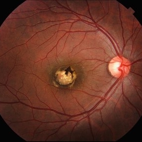
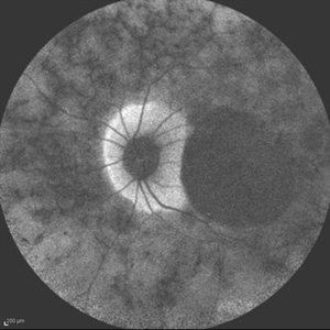
 Macular Coloboma (LE)
Sep 18 2024 by Anand Temkar
A 24 year old male came with chief complaint of diminution of vision in both eyes since childhood. Vision in both eyes was 6/24. IOP in RE was 12 and LE was 14 mm of Hg. On fundus examination periphery was within normal limits and central fundus revealed this picture. The serology testing such as serum IgM, IgG for toxoplasma and cytomegalovirus was negative. I have also uploaded LE color photo and BE OCT of this patient.
Photographer: Dr.Anand Temkar- Retina Foundation, Ahmedabad
Imaging device: Mirante
Condition/keywords: macular coloboma
-
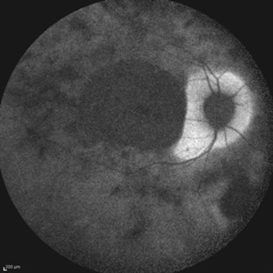
 Macular Coloboma (RE)
Sep 18 2024 by Anand Temkar
A 24 year old male came with chief complaint of diminution of vision in both eyes since childhood. Vision in both eyes was 6/24. IOP in RE was 12 and LE was 14 mm of Hg. On fundus examination periphery was within normal limits and central fundus revealed this picture. The serology testing such as serum IgM, IgG for toxoplasma and cytomegalovirus was negative. I have also uploaded LE color photo and BE OCT of this patient.
Photographer: Dr.Anand Temkar- Retina Foundation, Ahmedabad
Imaging device: Mirante
Condition/keywords: coloboma of macula
-
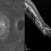
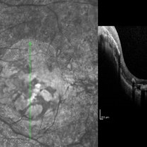 Macular Coloboma and Pigmentary Retinopathy
Feb 25 2017 by Hamid Ahmadieh, MD
Infrared and OCT images of the left eye of a 25-year-old woman with bilateral macular colobomata and pigmentary retinopathy similar to Leber's congenital amaurosis.
Photographer: Shabnam Poureh, Negah Eye Center, Tehran, Iran
Imaging device: Spectralis OCTc
Condition/keywords: infrared image
-
 Macular Coloboma and Pigmentary Retinopathy
Feb 25 2017 by Hamid Ahmadieh, MD
Infrared and OCT images of the left eye of a 25-year-old woman with bilateral macular colobomata and pigmentary retinopathy similar to Leber's congenital amaurosis.
Photographer: Shabnam Poureh, Negah Eye Center, Tehran, Iran
Condition/keywords: bilateral pigmentary retinopathy, infrared image, macular coloboma, optical coherence tomography (OCT)
-
 Macular Coloboma and Pigmentary Retinopathy
Feb 25 2017 by Hamid Ahmadieh, MD
Fundus autofluorescence (FAF) image of the left eye of a 25-year-old woman with bilateral macular colobomata and pigmentary retinopathy similar to Leber's congenital amaurosis.
Photographer: Shabnam Poureh, Negah Eye Center, Tehran, Iran
Condition/keywords: bilateral pigmentary retinopathy, fundus autofluorescence (FAF), macular coloboma
-
 Macular Coloboma and Pigmentary Retinopathy
Feb 25 2017 by Hamid Ahmadieh, MD
Infrared image of the left eye of a 25-year-old woman with bilateral macular colobomata and pigmentary retinopathy similar to Leber's congenital amaurosis.
Photographer: Shabnam Poureh, Negah Eye Center, Tehran, Iran
Condition/keywords: bilateral pigmentary retinopathy, infrared image, macular coloboma
-
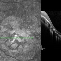
 Macular Coloboma and Pigmentary Retinopathy
Feb 25 2017 by Hamid Ahmadieh, MD
Infrared and OCT images of the right eye of a 25-year-old woman with bilateral macular colobomata and pigmentary retinopathy similar to Leber's congenital amaurosis.
Photographer: Shabnam Poureh, Negah Eye Center, Tehran, Iran
Imaging device: Spectralis OCT
Condition/keywords: infrared image, macular coloboma, optical coherence tomography (OCT)
-
 Macular Coloboma and Pigmentary Retinopathy
Feb 25 2017 by Hamid Ahmadieh, MD
Fundus autofluorescence (FAF) image of the right eye of a 25-year-old woman with bilateral macular colobomata and pigmentary retinopathy similar to Leber's congenital amaurosis.
Photographer: Shabnam Poureh, Negah Eye Center, Tehran, Iran
Condition/keywords: bilateral pigmentary retinopathy, fundus autofluorescence (FAF), macular coloboma
 Loading…