Search results (10 results)
-
 Fuchs' Heterochromic iridocyclitis
Mar 5 2021 by Niloofar Piri, MD
Heterochromia associated with Fuchs' heterochromic iridocyclitis in the left eye of a 63-year-old patient. Notice the green color of the left iris resulting from diffuse iris atrophy.
Photographer: Douglas Snyder, MD. St. Louis University
Condition/keywords: Fuchs, Fuchs' heterochromic cyclitis, heterochromia
-
---thumb.jpg/image-square;max$300,300.ImageHandler) Inferior Sector Iris Atrophy With Depigmentation
Inferior Sector Iris Atrophy With Depigmentation
Aug 1 2013 by From the Collections of Thomas M. Aaberg, MD and Thomas M. Aaberg Jr., MD
Inferior sector iris atrophy with depigmentation.
Condition/keywords: depigmentation, inferior sector iris atrophy
-
 Slide 12-10
Feb 27 2019 by Lancaster Course in Ophthalmology
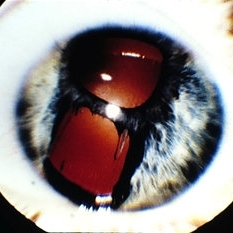
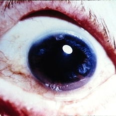
Essential iris atrophy. Pupil (above) and full-thickness iris hole show red reflex.
Condition/keywords: iris atrophy
-
 Slide 12-11
Feb 27 2019 by Lancaster Course in Ophthalmology
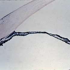
Essential iris atrophy. Anterior chamber angle closed by a peripheral anterior synechia (H&E x16).
Condition/keywords: iris atrophy
-
 Slide 12-12
Feb 27 2019 by Lancaster Course in Ophthalmology
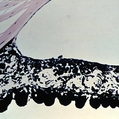
Essential iris atrophy. An area showing a full-thickness iris hole (H&Ex21).
Condition/keywords: iris atrophy
-
 Slide 12-13
Feb 27 2019 by Lancaster Course in Ophthalmology
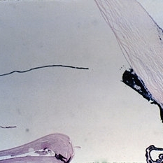
Essential iris atrophy. An area showing a full-thickness iris hole (H&Ex21).
Condition/keywords: iris atrophy
-
 Slide 12-7
Feb 27 2019 by Lancaster Course in Ophthalmology
Primary closed-angle glaucoma. The clinical triad of irregular pupil, segmental iris atrophy, and glaukomflecken is seen.
Condition/keywords: glaukomflecken, primary angle-closure glaucoma
-
 Slide 2-35
Feb 19 2019 by Lancaster Course in Ophthalmology
Atrophic iris in Fuch's heterochromic iridocyclitis. Mild plasma cell and lymphocytic infiltrate is present, as well as small vessels in the anterior border layer.
Condition/keywords: Fuchs' heterochromic cyclitis, iris atrophy, lymphocytes
-
 Slide 9-26
Feb 26 2019 by Lancaster Course in Ophthalmology
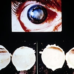
Hypotensive retinopathy. This patient presented with a slightly red eye and , aqueous cells, and flare. He developed areas of iris atrophy and cataract. Postmortem examination showed extensive cobblestone (paving-stone) degeneration which extended posteriorly to the equator in the right eye (lower left views). No cobblestone degeneration was present in the right eye (lower right views).
Condition/keywords: cataract, hypertensive retinopathy, iris, keratic precipitates
-
---thumb.JPG/image-square;max$300,300.ImageHandler) TID (Trans Illumination Defect)
TID (Trans Illumination Defect)
Jul 8 2013 by Jason S. Calhoun
74-year-old patient who VA 20/70 OD, 20/50 OS. Complaints of blurred vision. Iris atrophy, both eyes. ERM, right eye, Patient to have cataract surgery to improve distance vision.
Photographer: Jason S. Calhoun, Department of Ophthalmology, Mayo Clinic Jacksonville, Florida
Condition/keywords: translucency of iris
 Loading…