Search results (73 results)
-
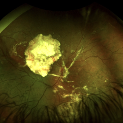
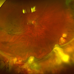
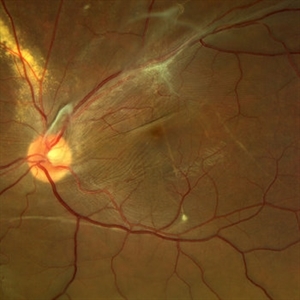 Displaced & folded macula
Oct 10 2022 by Ricardo Leitão Guerra
Tractional retinal detachment due to sickle cell retinopathy leading to a displaced and folded appearance of the macula in this 36-yo male. Subretinal bands are also noticed crossing the macula towards inferior retinal detachment area.
Photographer: Ricardo Leitão Guerra
Imaging device: Clarus 700 - Zeiss
Condition/keywords: folds, sickle cell retinopathy, subretinal bands, tractional retinal detachment
-
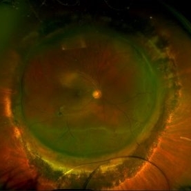
 Central Serous Retinopathy
Mar 19 2024 by Corey Grant
Ultra Wide-Field Fundus Autofluorescence Imaging of a 37 year old female with Central Serous Retinopathy affecting her right eye. Patient Visual Acuity was 20/20 in both eyes. Patient reported black spots in her vision onset three years ago, with associating flashes of light. Patient reports history of cortisone back injections a few years ago and denies Flonase use. The physician stated that there is hyperautofluorescence in the area of gutter of Sub-Retinal Fluid which likely happened from CSR.
Photographer: Corey Grant, OSC
Imaging device: OPTOS CALIFORNIA RGB
Condition/keywords: Central Serous Chorioretinopathy (CSR), central serous retinopathy (CSR), fundus autofluorescence (FAF), Guttering, hyperautofluorescence, inferior retina, OPTOS, Retina, Right Eye, subretinal fluid, ULTRA WIDE FIELD
-
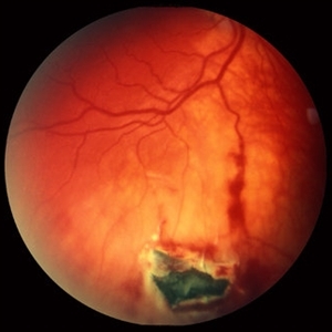
 Ozurdex Implant Related Tear
Jan 26 2022 by Tracey Grabowski
Ultra wide-field photograph of a 73-year-old female with an Ozurdex implant causing a retinal tear in the inferior retina. Prompt laser was added to prevent a retinal detachment and patient has been doing well since. Patient had no symptoms following the occurrence.
Photographer: Tracey Grabowski
Imaging device: Optos California
Condition/keywords: fundus photograph, inferior retina, optos, ozurdex, Ozurdex implant, retinal tear, treated retinal tear, ULTRA WIDE FIELD
-
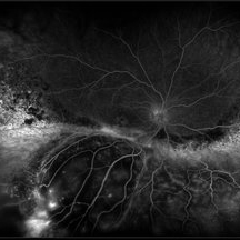
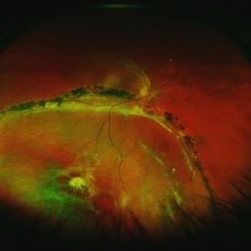
 Coats' Disease
Jul 16 2019 by Kim Barrett
Ultra-wide field fluorescein angiogram of a 23-year-old male with Coats' disease, presented with distorted vision affecting his left eye. He reported seeing flashes and floaters since January of 2019, but the flashes had resolved. He was treated with Intravitreal Preservative Free Triamcinolone in the office and scheduled for PRP laser in the near future.
Photographer: Kim Barrett
Imaging device: Optos
Condition/keywords: Coats' disease, fluorescein angiogram (FA), fluorescein leakage, inferior retina, ischemia, left eye, Optos, ultra-wide field imaging
-
 Dexamethasone Implant
Jul 3 2021 by Gerardo Rivera Arroyo
42-year-old male, operated on for vitrectomy plus scleral buckling plus silicone plus dexamethasone implant for inferior retinal detachment with PVR.
Photographer: Rosa Elizabeth Moreno Anda, MD, Hospital Central Militar CDMX
Condition/keywords: dexamethasone implant, retina surgery, vitrectomy
-
 Dislocated Crystalline Lens
Mar 19 2024 by Annaka Gooding
Ultra Wide field fundus photography of a 70 year old male who presented to clinic with a sudden increase of vision due to dropped crystalline lens secondary to severely dense cataract. Patient reported seeing a full black circle in his inferior visual field. Patient's visual acuity at time of visit was 20/100 with a +5.00 diopter lens. The physician recommended surgical intervention, and discussed surgery for PPV/PPL/IOL implantation with an ACIOL.
Photographer: Annaka Gooding, CPO
Imaging device: Optos California RGB
Condition/keywords: dislocated crystalline lens, fundus photography, inferior retina, OPTOS CALIFORNIA RGB, Right Eye, Ultra-wide field retinal imaging
-
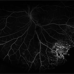
 Exudative Retinal Detachment and Branch Retinal Vein Occulsion
Oct 29 2020 by Olivia Rainey
Ultra-widefield fluorescein anigogram of a 51-year-old female with an exudative retinal detachment and branch retinal vein occlusion with retinal neovascularization affecting her right eye. The physician stated that the multiple aneurysmal dilations noted in the inferior periphery are responsible for the exudative RD seen on exam. He is considering Coat's vs FEVR given family history of aneurysms/congenital heart pathology per patient. He encouraged the patient to control their blood pressure, cholesterol, blood sugar, and co-morbidities which may have promoted the BRVO. He recommended antiVEGF injections to control the vascular leakage. Given the severe presentation and imminent threat to her vision, he recommended Eylea as first line therapy.
Photographer: Olivia Rainey, OCT-C, COA
Imaging device: Optos California
Condition/keywords: branch retinal vein occlusion (BRVO), chronic retinal detachment, fluorescein angiogram (FA), fluorescein leakage, inferior retina, inferior retinal detachment, Optos, ultra-wide field imaging
-
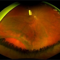
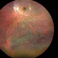 Inferior retinal detachment with lattice and holes
May 31 2023 by Aditya S Kelkar, MS, FRCS, FASRS,FRCOphth
Importance of dilated retina check up before Lasik surgery can't be better demonstrated...patient totally asymptomatic came for Lasik opinion and has inferior retinal detachment with lattice and holes, sparing the macula
Photographer: Dr. Sahil Wagh , National Institute of Opthalmology, Pune , India
Imaging device: Zeiss Clarus 500
Condition/keywords: inferior retinal detachment
-
 Methotrexate Bubble following Intravitreal Injection for PVR
Sep 21 2022 by Zach Seim
Ultra-widefield fundus photograph of an 81 year old female with a Methotrexate bubble following an Intravitreal Injection for Proliferative Vitreoretinopathy. Patient has been presenting to the office for two week interval Methotrexate injections in her left eye. The image was taken prior to her eighth injection which revealed a residual Methotrexate bubble in her inferior retinal image. This patient was seeing "lots" of floaters, as well as having visual acuity of cc20/400 cc20/200 PH.
Photographer: Zach Seim
Imaging device: OPTOS California
Condition/keywords: bubble, fundus photograph, fundus photography, intravitreal injection, left eye, methotrexate, nasal retina, Optos, proliferative vitreoretinopathy (PVR), pseudocolor, ultra-wide field imaging
-
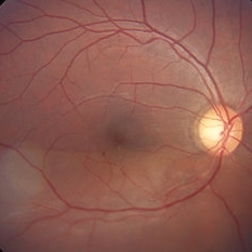
 Retinal detachment
Apr 12 2023 by Ahmed Abbas Hashmi, OD
Color fundus photograph of the left eye of a 30-year-old man with asymptomatic inferior retinal detachment with pigmented demarcation line. Macula and Disc healthy.
Photographer: Ahmed Abbas Hashmi
Imaging device: Topcon TRC-NW8F
Condition/keywords: Pigmentary demarcation line, Retinal Detachment
-
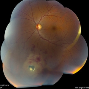
 Retinal Detachment After Retinoblastoma Treatment
Mar 10 2024 by Alexandre Grandinetti, MD, PhD
Inferior retinal detachment occurring 6 years after treatment with intraarterial chemotherapy and laser in an 8-year-old boy.
Photographer: Corina Szrek
Condition/keywords: pediatric, retinoblastoma
-
 Retinal Detachment Sparing Fovea By Microns
Sep 24 2018 by samarth mishra
A 29-year-old young female presented with complaint of blurring of vision in the right eye since one year. Best corrected visual acuity was 20/40. On routine examination inferior retinal detachment was noted. Optical coherence tomography (OCT) showed the retinal detachment sparing the fovea by few microns.
Photographer: Aditya Birla Sankara Nethralaya, Kolkata , West Bengal , India
Condition/keywords: color fundus photograph, multicolor, optical coherence tomography (OCT)
-
 Scleral Buckling IOL Drop
Aug 6 2023 by Dr.Sheetal Divate
A 27 year old female with an old history of trauma and operated with scleral buckling and cataract surgery in the past came recently with complaints of DOV . Findings noted where IOL drop, inferior retinal detachment and old scleral buckle indent.
Photographer: Dr.Sheetal Divate
Imaging device: Optos Advance
Condition/keywords: dislocated intraocular lens (IOL), Retinal Detachment, scleral buckle
-
 Intraocular Foreign Body, Metallic, in Inferior Retina with Hemorrhage
Oct 1 2012 by Jeffrey G. Gross, MD, FASRS
IOFB, metallic, in inferior retina with hemorrhage.
Condition/keywords: inferior retina, intraocular foreign body
-
 Acute Exudative Polymorphous Vitelliform Maculopathy Red Free OD
Aug 27 2014 by Flavio A. Rezende, MD, PhD
45-year-old man with mild decrease in vision after strong headache. Fundus showing multiple deep irregular vitelliform lesions spread throughout entire posterior pole OU, forming a typical level of subretinal confluent lesions at the inferior retinal vascular arcades. No primary tumor or metastasis found.
Photographer: Eduardo Martins, Pontifícia Universidade Católica - Rio de Janeiro, Brazil
Imaging device: Topcon TRC 50EX
Condition/keywords: polymorphous exudative vitelliform maculopathy
-
 Chronic Inferior Retinal Detachment
Mar 1 2017 by Philip J. Polkinghorne, MD
Color photograph of chronic retinal detachment with pigment demarcation line and atrophic holes visible. The vision was recorded at 20/20, and follow up is 3 years.
Photographer: Alex Fraser
Condition/keywords: atrophic retinal hole, demarcation line
-
 Hydrogel Implant Intrusion
May 5 2020 by Geovanni Jassiel Rios, MD
Ultra-wide field fundus photograph of the right eye with reattached retina. We can observe retinal hydrogel implant intrusion at the inferior retina
Photographer: Ericka , Hospital de la Luz
Condition/keywords: hydrogel implant intrusion, ultra-wide field imaging
-
 Old RRD With Retinal Cysts and High Watermark
Apr 10 2020 by Dipak Nag, MBBS, FCPS, MSc, FRF
Intra-operative fundus picture of a 20-year-old boy showing multiple retinal cysts and high watermark in a case of old inferior retinal detachment OD.
Photographer: Dipak
Condition/keywords: high watermark, retinal cyst
-
 Inferior retinal detachment
Dec 19 2012 by Eric A. Postel, MD
Color fundus photograph of an inferior retinal detachment
-
 Intraocular Foreign Body
Apr 9 2014 by Aleksandra V. Rachitskaya, MD, FASRS
Fundus photo of intraocular foreign body located in the inferior retina.
Photographer: Bascom Palmer Eye Institute
Condition/keywords: intraocular foreign body
-
 Retinal Detachment with Giant Tear
Mar 13 2018 by Olivia Rainey
Ultra-wide field pseduocolor image of a 36-year-old male with an giant inferior tear, causing a retinal detachment.
Photographer: Olivia Rainey
Imaging device: Optos
Condition/keywords: color fundus photograph, giant retinal tear, inferior retina, macular splitting, Optos, ultra-wide field imaging
-
 Acute Exudative Polymorphous Vitelliform Maculopathy Angio OD
Aug 27 2014 by Flavio A. Rezende, MD, PhD
45-year-old man with mild decrease in vision after strong headache. Fundus showing multiple deep irregular vitelliform lesions spread throughout entire posterior pole OU, forming a typical level of subretinal confluent lesions at the inferior retinal vascular arcades. No primary tumor or metastasis found.
Photographer: Eduardo Martins, Pontifícia Universidade Católica - Rio de Janeiro, Brazil
Imaging device: Topcon TRC 50EX
Condition/keywords: polymorphous exudative vitelliform maculopathy
-
 Acute Exudative Polymorphous Vitelliform Maculopathy Angio OS
Aug 27 2014 by Flavio A. Rezende, MD, PhD
45-year-old man with mild decrease in vision after strong headache. Fundus showing multiple deep irregular vitelliform lesions spread throughout entire posterior pole OU, forming a typical level of subretinal confluent lesions at the inferior retinal vascular arcades. No primary tumor or metastasis found.
Photographer: Eduardo Martins, Pontifícia Universidade Católica - Rio de Janeiro, Brazil
Imaging device: Topcon TRC 50EX
Condition/keywords: polymorphous exudative vitelliform maculopathy
-
 Acute Exudative Polymorphous Vitelliform Maculopathy Color OD
Aug 27 2014 by Flavio A. Rezende, MD, PhD
45-year-old man with mild decrease in vision after strong headache. Fundus showing multiple deep irregular vitelliform lesions spread throughout entire posterior pole OU, forming a typical level of subretinal confluent lesions at the inferior retinal vascular arcades. No primary tumor or metastasis found.
Photographer: Eduardo Martins, Pontifícia Universidade Católica - Rio de Janeiro, Brazil
Imaging device: Topcon TRC 50EX
Condition/keywords: polymorphous exudative vitelliform maculopathy
-
 Acute Exudative Polymorphous Vitelliform Maculopathy Color OS
Aug 27 2014 by Flavio A. Rezende, MD, PhD
45-year-old man with mild decrease in vision after strong headache. Fundus showing multiple deep irregular vitelliform lesions spread throughout entire posterior pole OU, forming a typical level of subretinal confluent lesions at the inferior retinal vascular arcades. No primary tumor or metastasis found.
Photographer: Eduardo Martins, Pontifícia Universidade Católica - Rio de Janeiro, Brazil
Imaging device: Topcon TRC 50EX
Condition/keywords: polymorphous exudative vitelliform maculopathy
 Loading…