Search results (76 results)
-
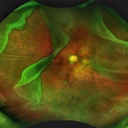
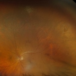
 A Vessel That Would Not Let Go
May 5 2025 by Malvika Singh
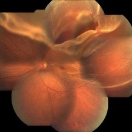
Fundus photograph of a retinal detachment showing a horse shoe shaped tear and a bridging vessel.
Photographer: Dr Tejaswita Verma, Retina Foundation, Ahmedabad, India
Imaging device: Mirante SLO/OCT
Condition/keywords: bridging vessel, horseshoe tear
-
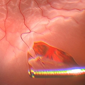
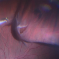 Intraoperative Photo Taken During Vitrectomy
Jan 26 2017 by Manish Nagpal, MD, FRCS (UK), FASRS
Intraoperative photo while doing vitectomy near a horseshoe tear to clear the adherent vitreous enhanced by peripheral scleral indentation while using chandelier light.
Photographer: Manish Nagpal
Imaging device: Still captured from a 3 chip HD camera on microscope
Condition/keywords: cutter, scleral indentation, vitrectomy, vitreous
-
 Retinal Detachment with Horseshoe Tear
Jun 10 2020 by Manish Nagpal, MD, FRCS (UK), FASRS
Localized superior retinal detachment with horseshoe tear and minimal fluid.
Photographer: gayathri mohan
Imaging device: nidek slo mirante
-
 Rhegmatogenous Retinal Detachment
Mar 3 2021 by Patrik Rajs
A 51-year-old female patient presented with inferior nasal scotoma and 5/10 vision in the right eye due to a retinal detachment with a giant retinal horseshoe tear.
Photographer: Patrik Rajs, EYE CLINIC of Jan Evangelista Purkyne University and Masaryk Hospital, Czech Republic, Ústí nad Labem
Imaging device: Clarus 700
Condition/keywords: giant retinal tear
-
 Giant Retinal Tear with Multiple Retinal Breaks
Apr 21 2025 by Hrishikesh Naik, MS
A 28 year old high myope with retinal detachment associated with a supero-temporal giant retinal tear in addition to multiple peripheral horseshoe tears and an additional supero-nasal retinal tear.
Photographer: Hrishikesh Naik
Imaging device: Optos Daytona
Condition/keywords: giant retinal tear, High Myopia, horseshoe tear, retinal break, retinal detachment
-
 Horseshoe Tear in Retinitis Pigmentosa
Mar 22 2021 by ASRS Staff
Montage of 25-year-old patient, high myopic patient came with complaint of diminution of vision in both eyes and on posterior segment examination of right eye, HST was present along with maculopathy.
Imaging device: Nidek Mirante
Condition/keywords: maculopathy, retinitis pigmentosa
-
 Hosreshoe Tears on Posterior Pole
Mar 22 2025 by Deepak Bhojwani, MS
A fundus image of an asymptomatic 64 year old male with large horseshoe shaped breaks in inferonasal quadrant on posterior pole, an unusual location for retinal breaks.
Photographer: DR DEEPAK BHOJWANI
Condition/keywords: horseshoe tear, posterior pole break, retinal break
-
 Rhegmatogenous Retinal Detachment
Jul 14 2016 by PAVEL FLORES-MORENO
Superior horseshoe tear with rhegmatogenous retinal detachment macula off.
Photographer: Pavel Flores-Moreno
Imaging device: Smartphone
-
 Sub Retinal Gas 1 Day Post Pneumatic Retinopexy
Jul 8 2016 by Asaf Friehmann
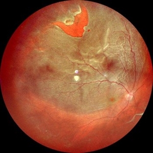
Fundus photograph of an 71-year-old male with a sub retinal C3F8 1 day after pneumatic retinopexy for the treatment of rhegmatogenous retinal detachment involving a single 1 hour horseshoe tear at 12 o'clock.
Photographer: Lilach Gorek
Condition/keywords: pneumatic retinopexy
-
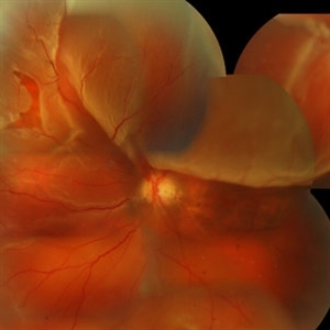 Superior Bullous Detachment With Horseshoe Tear
May 15 2014 by Manish Nagpal, MD, FRCS (UK), FASRS
Patient having bullous superior retinal detachment with a horseshoe tear.
Photographer: pooja barot, Optometrist, Retina Foundation, Ahmedabad
-
 Macula on Retinal Detachment with large Horseshoe Tear
Apr 26 2023 by Kelli Nyenhuis
Optos photograph of a 61-year-old male with a macula on retinal detachment and large horseshoe tear. Patient had no visual changes.
Photographer: Kelli Nyenhuis, COA
Imaging device: Optos California
-
 Rhegmatogenous Retinal Detachment
Jan 8 2019 by ANUBHAV GOYAL, DNB, FVRS
Fresh rhegmatogenous retinal detachment with big posterior horseshoe tear.
Photographer: Anubhav Goyal, Giridhar eye institute, cochin, India
-
 The Horseshoe Of Havoc
Jun 28 2025 by Tejaswita Verma
Fundus image of a 50 year old male with a very large horseshoe tear causing RRD with macula off, hydration folds.
Photographer: Dr. Tejaswita Verma
Imaging device: MIRANTE
Condition/keywords: horseshoe tear, large break
-
 Acute Retinal Detachment
Nov 9 2012 by Norman Byer
This 54-year-old man was referred because of sudden symptoms in his opposite eye in which he had suffered an acute retinal detachment secondary to a horseshoe tear around lattice degeneration. During the examination, the fellow eye shown here was also found to have this large horseshoe tear about 1 o’clock hour (4 disc diameters) in size. A tear occurred around a lattice lesion which is present on the flap but is out of focus. This tear had been asymptomatic even though it was caused by a posterior vitreous detachment and illustrates that even very large tears may produce no symptoms or mild symptoms that are easily overlooked.
Condition/keywords: lattice degeneration, posterior vitreous detachment
-
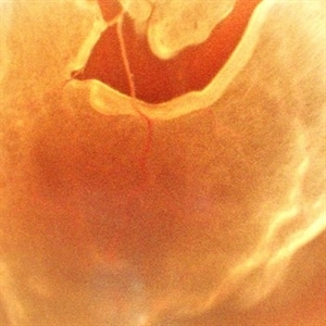
 Bleeding Bridging Vessel
Mar 27 2018 by Alan Sheyman, MD
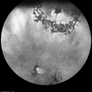
This is an infrared reflectance image of a horseshoe tear previously surrounded by laser retinopexy with a bridging vessel beautifully visible now causing recurrent vitreous hemorrhage.
Photographer: Karen Klima, University of Maryland
Imaging device: Heidelberg Spectralis
Condition/keywords: retinal tear, vitreous hemorrhage
-
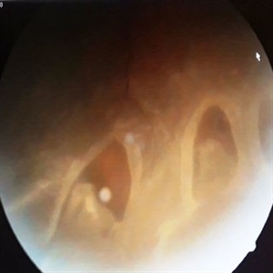
 Endoscopy: Open Angle Glaucoma and Recurrent Retinal Detachment
Dec 10 2012 by Yale L. Fisher, MD
Endoscopically controlled surgical repair of a rhegmatogenous retinal detachment in a phakic glaucoma patient with a fixed miotic pupil. The formed vitreous is still attached in the posterior pole but separated peripherally. A horseshoe tear is located in the superior nasal portion of the globe. The flap is trimmed and the posterior formed vitreous face is removed from the optic nerve and posterior pole with an open vertical scissors blade. An air fluid exchange through the break completes the reattachment.
Condition/keywords: video
-
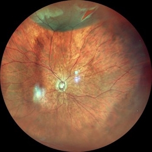
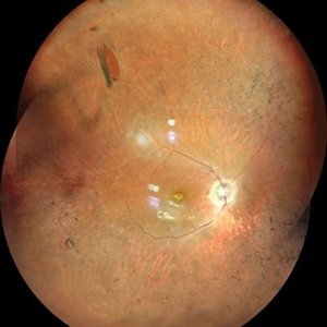
 ERMageddon - Wrinkle in the Space-time Fabric of Macula
Oct 29 2025 by SHRADDHA RAJ SHRIVASTAVA
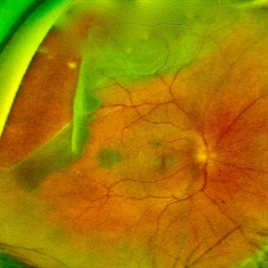
38 year old female with Epiretinal Membrane (ERM) over macula, post laser barrage for multiple symptomatic Horse-shoe Tears (HSTs) and Lattice Degenerations. Posterior pole revealed tilted disc with peripapillary atrophy. There is thick opaque epiretinal membrane obscuring the underlying superior arcade vessels and causing foveal ectopia with distortion of perimacular vasculature. Patient was planned for Right Eye pars plana vitrectomy for ERM peeling.
Photographer: Dr. Shraddha Raj Shrivastava
Imaging device: Nidek Mirante SLO/OCT (Confocal scanning/Spectral domain OCT
Condition/keywords: ectopic fovea, epiretinal membrane (ERM), ERM, horseshoe tear, vitreomacular traction (VMT)
-
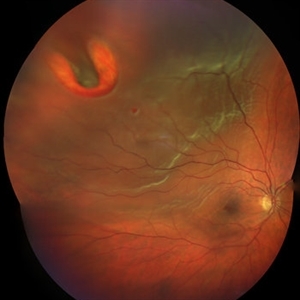
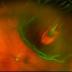
 ERMageddon - Wrinkle in the Space-time Fabric of Macula
Oct 29 2025 by SHRADDHA RAJ SHRIVASTAVA
38 year old female with Epiretinal Membrane (ERM) over macula, post laser barrage for multiple symptomatic Horse-shoe Tears (HSTs) and Lattice Degenerations (seen on wide-field image). Posterior pole revealed tilted disc with peripapillary atrophy. There is thick opaque epiretinal membrane obscuring the underlying superior arcade vessels and causing foveal ectopia with distortion of perimacular vasculature. Patient was planned for Right Eye pars plana vitrectomy for ERM peeling.
Photographer: Dr. Shraddha Raj Shrivastava
Imaging device: Nidek Mirante SLO/OCT (Confocal scanning/Spectral domain OCT
Condition/keywords: BARRAGE LASER, ectopic fovea, epiretinal membrane (ERM), horseshoe tear, lattice degeneration, vitreomacular traction (VMT)
-
 Giant Retinal Tear
Apr 29 2021 by Fong May Chew, FRCOphth, MBBS, BSc
Optos pictures of a 56-year-old man who presented with a giant retinal tear who extending from 7-12 o clock with a separate horseshoe tear at 1 o clock. Treated with pneumatic retinopexy.
Photographer: Hesham Hamli
Condition/keywords: giant retinal tear, pneumatic retinopexy
-
 Horse Shoe Tear With Retinal Detachment
Apr 28 2025 by rohan jain
56 year-old male with idiopathic HST and RRD
Photographer: Dr. ROHAN JAIN
Condition/keywords: horseshoe tear, Retinal Detachment, rrd
-
 Horseshoe Retinal Tear
Aug 6 2025 by Korey Starkey
80 year-old patient presented with HSRT without detachment in the left eye and macula-off detachment in the right eye. Scheduled patient for prompt surgical repair OD and same day laser retinopexy OS to reduce risk of retinal detachment.
Photographer: Korey Starkey
Imaging device: Optos
Condition/keywords: color fundus photograph, fundus photography, horseshoe tear, Optos
-
 Horseshoe Tear
Jun 24 2015 by Andree Henaine-Berra, MD
Photograph of the right eye of a 58-year-old male patient with a retinal detachment due to a peripheral horseshoe tear, showing the moment when cryotherapy is applied during the scleral bluckling procedure.
Photographer: Jorge Morales, MD. Hospital General "Dr. Manuel Gea Gonzalez". Mexico City.
Condition/keywords: acute retinal detachment, cryotherapy, scleral buckle
-
 Horseshoe Tear
Jun 16 2024 by Anjana Mirajkar, MS Ophthalmology
An intra operative image showing a large horse shoe tear with retinal detachment.
Photographer: Dr. Anjana Mirajkar -Retina Foundation, Ahmedabad
Condition/keywords: rhegmatogenous retinal detachment
-
 Horseshoe Tear
Apr 9 2017 by Aliya Sultana
Fundus photograph of an 41-year-old man with multiple horseshoe tears in the retina, patient is myopic with -3.00 Dsph in both eyes. Suddenly patient noticed loss of vision in left eye , presented to our department next day. Other eye showed lattice degeneration . Patient underwent pars plana vitrectomy with silicone oil tamponade.
Photographer: Dr Aliya Sultana , Assistant Professor,Sarojini Devi Eye Hospital, Hyderabad, Telangana. India.
Condition/keywords: myopia
-
 Horseshoe Tear
Sep 17 2015 by Jason S. Calhoun
Horseshoe tear with sub retinal fluid present superior temporal in the left eye.
Photographer: Jason Calhoun, Mayo Clinic, Department of Ophthalmology
Imaging device: OPTOS 200TX
 Loading…