Search results (5 results)
-
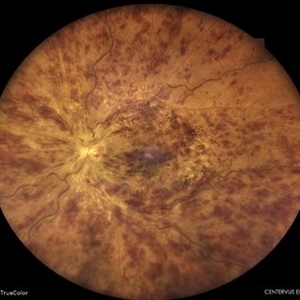 Central Retinal Vein Occlusion
Feb 25 2025 by Prithvi Chandrakanth
A 61-year-old woman with a history of hypertension noticed a sudden painless blurring of vision in her left eye. Over the next few days, the blurriness persisted, and she experienced a mild central scotoma. On examination, Fundoscopic evaluation revealed dilated, tortuous retinal veins, retinal hemorrhages, and macular oedema.
Photographer: DR.PRITHVI CHANDRAKANTH, DR.CHANDRAKANTH NETHRALAYA, KOZHIKODE
Imaging device: EIDON
Condition/keywords: CRVO, CRVO WITH MACULA EDEMA, flame shaped retinal hemorrhage
-
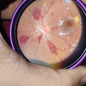 Thrombocytopenia
Sep 24 2024 by DR Rohit Gupta
Fundus photography of a 16 year-old girl suffering from severe thrombocytopenia, showing flame shaped hemorrhage.
Photographer: Dr Rohit gupta
Imaging device: Samsung S21
Condition/keywords: anaemic retinopathy, flame shaped retinal hemorrhage, Haemorrhage, Roth spots, white centered retinal hemorrhage (Roth Spot), white dot syndrome
-
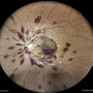 Anaemic Retinopathy
May 8 2023 by Akansha Sharma
Colour fundus photograph of a 38 year old male with anaemic retinoathy
Photographer: Dr. Urmil Shah, Dr. Denish Patel, Dr. Akansha Sharma, Bharati Eye Clinic, Ahmedabad, Gujarat
Condition/keywords: anaemic retinopathy, flame shaped retinal hemorrhage, subhyaloid hemorrhage
-
---thumb.jpg/image-square;max$300,300.ImageHandler) Diabetes with Flame Hemorrhage FA
Diabetes with Flame Hemorrhage FA
Apr 4 2014 by H. Michael Lambert, MD
Diabetes.
Photographer: Donald Lowd
Condition/keywords: diabetes, flame shaped retinal hemorrhage
-
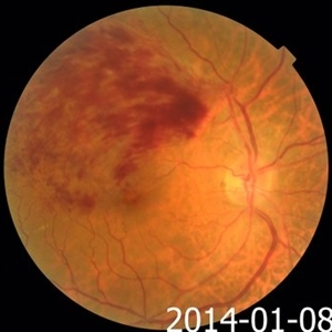 Recurrent Branch Retinal Vein Occlusion
May 31 2014 by Hamid Ahmadieh, MD
Color fundus photograph of the right eye of a 60-year-old man with recurrent BRVO.
Photographer: Soodabeh Fooladin, Negah Eye Center, Tehran, Iran
Condition/keywords: branch retinal vein occlusion (BRVO), color fundus photograph, flame shaped retinal hemorrhage
 Loading…