Search results (15 results)
-
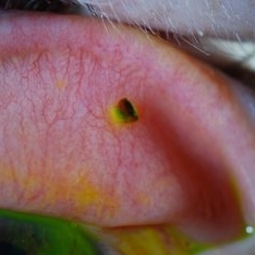
 Corneal Abrasion With Foreign Body Present
Jul 8 2013 by Jason S. Calhoun
ER patient came in with corneal abrasion in the left eye which was getting worst. VA 20/25. Slit lamp exam showed corneal abrasion superiorly at 2-o'clock. Flipped eyelid and foreign body appeared on the upper tarsal plate. Foreign body was removed.
Photographer: Jason S. Calhoun, Department of Ophthalmology, Mayo Clinic Jacksonville, Florida
Condition/keywords: intraocular foreign body, perforated corneal ulcer
-
 Corneal Ulcer
Sep 14 2017 by Theodore Leng, MD, MS, FASRS
Corneal ulcer with hypopyon
Condition/keywords: central corneal ulcer, hypopyon
-
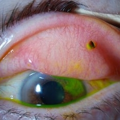
 Herpes Simplex
Sep 24 2024 by DR Rohit Gupta
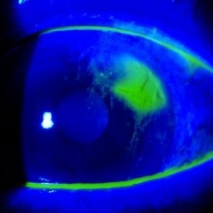
Slit lamp photograph of a 32 year-old male presented with redness, photophobia, and pain in left eye.
Photographer: Dr Rohit gupta
Imaging device: Samsung S21
Condition/keywords: corneal ulcer, Herpes, herpes dendrite, Herpes simplex infection
-
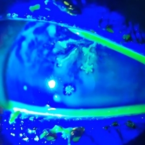 Herpetic Corneal Ulcer
Sep 24 2024 by DR Rohit Gupta
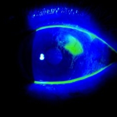
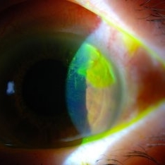
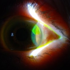
Slit lamp photograph of 32 year old male presented with herpetic corneal ulcer on staining with fluorescein dye under cobalt blue filted dendrits can be seen.
Photographer: Dr Rohit gupta
Imaging device: Samsung S21
Condition/keywords: corneal ulcer, dendritic keratitis, herpes dendrite, Herpes simplex infection, Herpes zoster, staining
-
 Hole in Cornea
Jul 14 2013 by Jason S. Calhoun
Slit lamp photo shows central hole which aqueous humor was leaking out.
Photographer: Jason S. Calhoun, Department of Ophthalmology, Mayo Clinic Jacksonville, Florida
Imaging device: TOPCON D-90 SL NIKON CAMERA
Condition/keywords: central corneal ulcer
-
---thumb.JPG/image-square;max$300,300.ImageHandler) Hole in Cornea
Hole in Cornea
Jul 14 2013 by Jason S. Calhoun
Slit lamp photo shows central hole which aqueous humor was leaking out.
Photographer: Jason S. Calhoun, Department of Ophthalmology, Mayo Clinic Jacksonville, Florida
Imaging device: TOPCON D-90 SL NIKON CAMERA
Condition/keywords: central corneal ulcer
-
---thumb.JPG/image-square;max$300,300.ImageHandler) Hole in Cornea 1
Hole in Cornea 1
Jul 14 2013 by Jason S. Calhoun
Slit lamp photo shows central hole which aqueous humor was leaking out.
Photographer: Jason S. Calhoun, Department of Ophthalmology, Mayo Clinic Jacksonville, Florida
Imaging device: TOPCON D-90 SL NIKON CAMERA
Condition/keywords: central corneal ulcer
-
 Prolapsed Iris
Jul 14 2013 by Jason S. Calhoun
Corneal ulcer developed into prolapsed iris.
Photographer: Jason S. Calhoun, Department of Ophthalmology, Mayo Clinic Jacksonville, Florida
Imaging device: TOPCON D-90 SL NIKON CAMERA
Condition/keywords: prolapse of iris
-
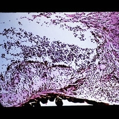
 Slide 1-19
Feb 19 2019 by Lancaster Course in Ophthalmology
Large "mutton-fat" keratic precipitates composed of macrophages, lymphoeytes, and epithelioid cells in a granulomatous corneal ulcer. (H&E stain)
Condition/keywords: corneal ulcer, epithelioid cells, lymphocytes, macrophages
-
 Slide 1-5
Feb 19 2019 by Lancaster Course in Ophthalmology
Pus (hypopyon) in the angle of the anterior chamber, secondary to a bactecorneal ulcer. (H&E stain)
Condition/keywords: bactecorneal ulcer, hypopyon, pus
 Loading…