Search results (65 results)
-
 Retinal Detachment Associated with Coloboma
Aug 23 2020 by Noy Ashkenazy, MD, MS
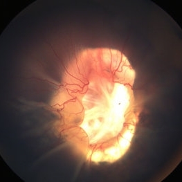
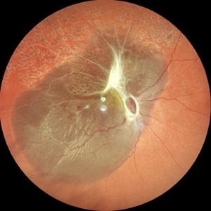
Fundus photograph of a 2-year-old boy with a history of CHARGE syndrome. The image nicely illustrates a retinal detachment associated with a congenital coloboma.
Photographer: Giselle DeOliveira
Imaging device: Retcam III
Condition/keywords: CHARGE syndrome, chronic retinal detachment, coloboma, pediatric retina
-
 Fundus Photo of Closed Funnel Retinal Detachment
Apr 10 2024 by Max D Schlesinger, MD
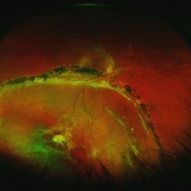
Wide-field funds photography of a closed funnel retinal detachment; patient had previously undergone 360 degree retinectomy in attempt to re-attach retina for a chronic retinal detachment, which was unsuccessful.
Condition/keywords: Closed funnel RD, detachment, Optos
-
 Synchysis Scintillans
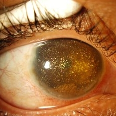
Sep 17 2015 by Jessica G Lee, MD
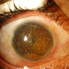
24-year-old male with history of chronic retinal detachment.
Photographer: Bob Masini
Condition/keywords: cholesterol crystals, refractile bodies, synchysis scintillans, trauma, vitreous hemorrhage
-
 Chronic Open Funnel Retinal Detachment With Horse Shoe Tear
Feb 7 2024 by Harsh Vardhan Singh, MS
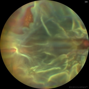
67 year old male with history of cataract surgery 1 year presented with old chronic retinal detachment with open funnel configuration with multiple breaks.
Photographer: Harsh Vardhan Singh
Imaging device: Clarus 700
Condition/keywords: chronic retinal detachment, Retinal Detachment, Retinal Detachment with multiple breaks
-
 Exudative Retinal Detachment and Branch Retinal Vein Occulsion
Oct 29 2020 by Olivia Rainey
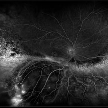
Ultra-widefield fluorescein anigogram of a 51-year-old female with an exudative retinal detachment and branch retinal vein occlusion with retinal neovascularization affecting her right eye. The physician stated that the multiple aneurysmal dilations noted in the inferior periphery are responsible for the exudative RD seen on exam. He is considering Coat's vs FEVR given family history of aneurysms/congenital heart pathology per patient. He encouraged the patient to control their blood pressure, cholesterol, blood sugar, and co-morbidities which may have promoted the BRVO. He recommended antiVEGF injections to control the vascular leakage. Given the severe presentation and imminent threat to her vision, he recommended Eylea as first line therapy.
Photographer: Olivia Rainey, OCT-C, COA
Imaging device: Optos California
Condition/keywords: branch retinal vein occlusion (BRVO), chronic retinal detachment, fluorescein angiogram (FA), fluorescein leakage, inferior retina, inferior retinal detachment, Optos, ultra-wide field imaging
-
 IOL
IOL
Jan 17 2018 by Emily Cooper
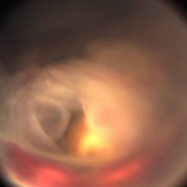
Optos image of 47-year-old man with a now worsening retinal detachment that had been treated by pneumatic retinopexy.
Photographer: Emily Cooper, Retina Specialists of Michigan
Imaging device: Optos Ultra Wide Field
Condition/keywords: chronic retinal detachment, intraocular lens (IOL)
-
 Macrocyst in the Fovea
Feb 2 2021 by Peter J Belin, MD
36-year-old male with a white cataract and a chronic total retinal detachment for 1 year presented with a recurrent PVR detachment after primary repair 2 weeks prior. This OCT- EDI demonstrates a large retinal cyst through the fovea.
Photographer: Holly Cheshier, CRA, OCT-C, COT
Imaging device: Heidelberg Spectralis
Condition/keywords: chronic retinal detachment, proliferative vitreoretinopathy (PVR), retinal cyst, retinal macrocyst
-
 Macrocysts in Kickboxer
Nov 17 2023 by Bradley T. Smith, MD, FASRS
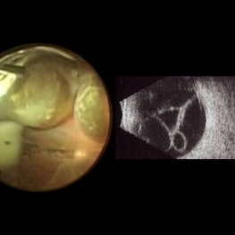
Intraoperative photo and preoperative b scan of chronic retinal detachment with macrocysts in a kickboxer
Condition/keywords: B scan ultrasound, chronic retinal detachment, ocular trauma, pars plana vitrectomy (PPV), retinal macrocyst
-
 Retinal Cyst
Aug 14 2020 by Noy Ashkenazy, MD, MS
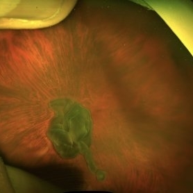
Fundus photograph of a 13-year-old male with a chronic retinal detachment following a penetrating ocular trauma. There is a retinal cyst and proliferative vitreoretinopathy.
Photographer: Giselle DeOliveira
Imaging device: Retcam III
Condition/keywords: chronic retinal detachment, proliferative vitreoretinopathy (PVR), retinal cyst
-
 Synchisis Scintillans
Sep 17 2015 by Jessica G Lee, MD
24-year-old male with history of chronic retinal detachment.
Condition/keywords: cholesterol crystals, refractile bodies, synchysis scintillans, trauma, vitreous hemorrhage
-
 The Bullet Ridden Retina
Feb 17 2024 by SHISHIR VERGHESE, MS, FVRS, FAICO (Retina)
Fundus image obtained of a case of lasered branch retinal vein occlusion (BRVO) with fibrovascular proliferation (FVP) where the laser marks have given way to multiple small retinal holes due to traction from the same.
Photographer: DIVYA SHAJI
Imaging device: NIDEK MIRANTE
Condition/keywords: BRVO, chronic retinal detachment
-
 Chronic Inferior Retinal Detachment
Mar 1 2017 by Philip J. Polkinghorne, MD
Color photograph of chronic retinal detachment with pigment demarcation line and atrophic holes visible. The vision was recorded at 20/20, and follow up is 3 years.
Photographer: Alex Fraser
Condition/keywords: atrophic retinal hole, demarcation line
-
 Intraretinal Cysts in Chronic Retinal Detachment
Dec 8 2020 by Alice Kim
B-scan ultrasound showing multiple intraretinal cysts in the setting of chronic retinal detachment and proliferative vitreoretinopathy.
Condition/keywords: chronic retinal detachment, intraretinal cyst, proliferative vitreoretinopathy (PVR)
-
 Chronic Retinal Detachment
Oct 12 2012 by Jeffrey G. Gross, MD, FASRS
Chronic retinal detachment, with precipitates.
Condition/keywords: chronic retinal detachment, precipitates
-
 Chronic Retinal Detachment
Oct 12 2012 by Jeffrey G. Gross, MD, FASRS
Chronic RD with multiple retinal cysts, B scan ultrasound.
Condition/keywords: B scan ultrasound, chronic retinal detachment, retinal cyst
-
 Angioma from Chronic Retinal Detachment
Sep 19 2014 by David Callanan, MD
36-year-old female, angioma from chronic retinal detachment.
Condition/keywords: angioma
-
 Angioma from Chronic Retinal Detachment
Sep 19 2014 by David Callanan, MD
36-year-old female, angioma from chronic retinal detachment.
Condition/keywords: angioma
-
 Angioma from Chronic Retinal Detachment
Sep 19 2014 by David Callanan, MD
36-year-old female, angioma from chronic retinal detachment.
Condition/keywords: angioma
-
 Angioma from Chronic Retinal Detachment
Sep 19 2014 by David Callanan, MD
36-year-old female, angioma from chronic retinal detachment.
Condition/keywords: angioma
-
 Angioma from Chronic Retinal Detachment
Sep 19 2014 by David Callanan, MD
36-year-old female, angioma from chronic retinal detachment.
Condition/keywords: angioma
-
 Angioma from Chronic Retinal Detachment
Sep 19 2014 by David Callanan, MD
36-year-old female, angioma from chronic retinal detachment.
Condition/keywords: angioma
-
 Angioma from Chronic Retinal Detachment
Sep 19 2014 by David Callanan, MD
36-year-old female, angioma from chronic retinal detachment.
Condition/keywords: angioma
-
 Angioma from Chronic Retinal Detachment
Sep 19 2014 by David Callanan, MD
36-year-old female, angioma from chronic retinal detachment.
Condition/keywords: angioma
-
 Angioma from Chronic Retinal Detachment
Sep 19 2014 by David Callanan, MD
36-year-old female, angioma from chronic retinal detachment.
Condition/keywords: angioma
-
 Angioma from Chronic Retinal Detachment
Sep 19 2014 by David Callanan, MD
36-year-old female, angioma from chronic retinal detachment.
Condition/keywords: angioma
 Loading…