Search results (83 results)
-
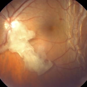
 Choroidal Detachment
Jan 17 2022 by Logan ryzenga
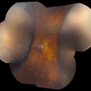
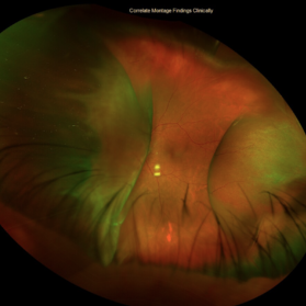
Left ultra-wide field photograph of an 81-year old female with a choroidal detachment affecting her left eye. Patient had a stent placed November, 2021 and following the procedure she complains of variable blurred vision and severe constricted visual fields. She presented at our office with flashes a month prior but without pain or floaters.
Photographer: Logan Ryzenga
Imaging device: Optos California
Condition/keywords: choroidal detachment, fundus photograph, left eye, Optos, pseudocolor, superior retina, ultra-wide field imaging
-
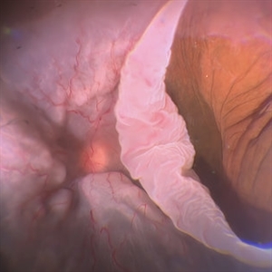 Giant Retinal Tear with Choroidal Detachment
Jun 12 2024 by Anand Temkar
Intra operative still of a 34 year old male showing Giant Retinal Tear with Choroidal Detachment.
Photographer: Dr.Anand Temkar- Retina Foundation, Ahmedabad
Condition/keywords: choroidal detachment, giant retinal tear
-
 Kissing Serous Choroidal Detachment
Mar 8 2023 by Annaka Gooding
Ultra-widefield fundus photograph of a 73 year old male with a Kissing serous choroidal detachment affecting his right eye. Patient presented at the office following a XEN implant and his vision was sc20/100 PH20/50+1. The physician recommended to start Prednisone treatment.
Photographer: Annaka Gooding
Imaging device: Optos California
Condition/keywords: fundus photography, Kissing Serous Choroidal Detachment, Optos, Right Eye, ultra-wide field imaging
-
 Retained Lens Fragment
Mar 2 2014 by Homayoun Tabandeh, MD, FASRS
Retained lens fragment, choroidal detachment, and serous retinal detachment post cataract surgery
Condition/keywords: retained lens fragments
-
 Choroidal and Retinal Detachment Secondary to Full-thickness Macular Hole
May 24 2024 by Tony Y Chen, MD
Optos photograph of a 61-year-old woman with choroidal and retinal detachment secondary to full-thickness macular hole.
Condition/keywords: choroidal detachment, Retinal detachment with macular hole
-
 Choroidal Detachment
Oct 4 2018 by Emily Cooper
Optos photograph of an 80-year-old man presenting with red, painful eye after heart surgery.
Photographer: Emily Cooper, Retina Specialists of Michigan, Grand Rapids MI
Imaging device: Optos
Condition/keywords: choroidal detachment, posterior scleritis
-
 Hemorrhagic Choroidals
Jan 22 2025 by Danish Shabbir, Ophthalmic Technologist
78 year old female complains of suddenly vision decrease 2 days ago.
Photographer: Danish Shabbir,Retina-EyeCare Centre
Imaging device: Optos California
Condition/keywords: choroidal detachment, Retinal Detachment, retinal detachment with choroidal
-
 Three Kisses
Mar 18 2025 by Gustavo Uriel Fonseca Aguirre
Cross-section of a B-mode ultrasound showing a kiss-shaped choroidal detachment; three lobes, giving the appearance of three kisses.
Photographer: Gustavo U. Fonseca Aguirre, Hospital Conde de Valenciana, Ciudad de México
Condition/keywords: Kissing Choroidal Detachment
-
 Uveal Effusion Syndrome
Oct 23 2023 by Gustavo Aguirre-Suarez
Fundus photograph of a 58-year-old man with Type 1 Uveal Effusion Syndrome, showing 360º bullous choroidal detachment.
Photographer: Dr. Gustavo Aguirre-Suarez
Imaging device: Zeiss Clarus 700
Condition/keywords: choroidal effusion, idiopathic uveal effusion syndrome
-
 Choroidal Detachment OS
Dec 2 2019 by Kristen Wagner
Choroidal Detachment of the left eye. Patient's vision was Hand Motion best corrected.
Photographer: Kristen Wagner, COT, OSC, Ophthalmic Photographer, Tennessee Retina
Condition/keywords: choroidal detachment
-
 Choroidal Detachment
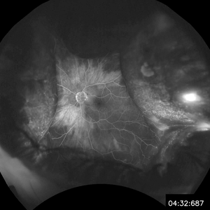
Jan 6 2020 by Sarah Oelrich
Choroidal detachment
Photographer: Sarah Oelrich CRA COT OCT-C
Imaging device: Optos
Condition/keywords: choroidal detachment, detachment, FA late phase
-
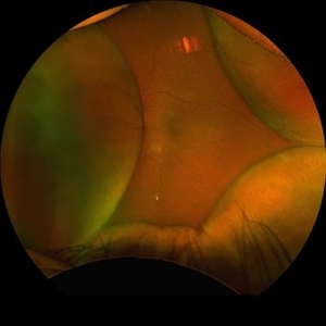
 Non-Kissing Choroidal Detachment
Apr 3 2023 by Zach Seim
An ultra-widefield fundus image of an 85 year old female with a Choroidal Detachment. Patient's vision at the time of the image was HM and surgery was not recommended.
Photographer: Zach Seim
Imaging device: Optos California
Condition/keywords: choroid, choroidal detachment, left eye, OPTOS CALIFORNIA, ultra-widefield image
-
 Ocular Hypotony Due to Leaking Bleb
Apr 1 2019 by Anfisa Ayalon, MD
81-year-old male who had trabeculectomy in his right eye 4 years ago, presented to the emergency room with complains of decreased vision in that eye for two months. Slit-lamp examination showed cystic bleb with leakage, intraocular pressure was 0 MMHg. Fundus examination showed hypotony maculopathy, peripheral choroidal detachments, multiple chorioretinal folds with subretinal fluid.
Photographer: Anfisa Ayalon, MD., Meir Medical Center, Kfar Saba, Israel.
Imaging device: California, Optos 200 DTX
Condition/keywords: choroidal detachment, hypotonous retinopathy, hypotony maculopathy
-
 24 Hours Post Scleral Wound Closure+ Scleral Buckle+25 g Vitrectomy+Silicon Oil
Jan 23 2015 by Carlos Quezada-Ruiz, MD, FASRS
24 hours post op fundus photograph of a 43-year-old man who had perforating injury to the right eye with a small piece of plastic while he was hammering. OD LP, subconjunctival hemorrhage, clear cornea, hyphema, irido and ciclodyalisis as well as a luxated lens with traumatic cataract and a dense vitreous hemorrhage. B-US showed rhegmatogenous retinal detachment with a tear and a big inferior hemorrhagic choroidal detachment. 360 peritomy revealed 2-entry scleral wounds were found in zone II (M V and M VI) and closure was performed. 25 G PPV was performed with the infusion canal placed in the AC through the limbus. Lensectomy and removal of a dense recent vitreous hemorrhage revealed a white detached retina with an exit wound through the temporal inferior segment of the optic nerve with a nasal GRT and sub retinal hemorrhage as well as temporal inferior choroidal, PVD was induced and PFOs helped stabilizing the retina while vitrectomy and sub-retinal hemorrhage was removed through the GRT. Fluid air exchange was made and 360 endolaser over the buckle indentation was done and silicon oil was used as endotamponade. This picture was taken 24 hrs after the surgery.
Photographer: Lilibeth Rodriguez, Instituto de la Visión. Torreon, Mexico.
Condition/keywords: central retinal artery occlusion (CRAO), giant retinal tear, trauma
-
 Choroidal Detachment
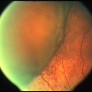
Mar 29 2013 by Henry J. Kaplan, MD
One quadrant choroidal detachment as brownish convex lesion.
Condition/keywords: choroidal detachment
-
 Choroidal Detachment, In Stereo
Sep 25 2012 by Michael P. Kelly, FOPS
Photographer: Michael P. Kelly, FOPS Director, Duke Eye Labs, Duke University Hospital, Duke Eye Center, Durham, NC
Imaging device: Zeiss FF3C
Condition/keywords: choroidal detachment, stereo pair
-
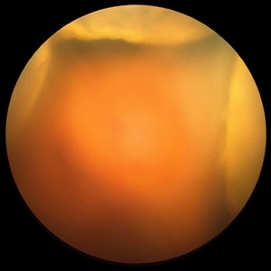
360 Choroidal Detachment in Uveal Effusion Syndrome
Apr 11 2025 by Siri Uppuluri
Fundus photograph of a phakic left eye in an 82-year-old man demonstrating 360 choroidal detachment secondary to uveal effusion syndrome. He underwent sclerectomy and drainage of the choroidal effusions with resolution after surgical intervention.
Photographer: Siri Uppuluri, MD; Rutgers New Jersey Medical School
Condition/keywords: choroidal detachment
-
 360 Degrees Choroidal Detachment
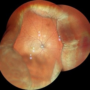
Sep 22 2024 by Anand Temkar
A 52 year old male came with chief complaints of diminution of vision in RE since past 15 days. He gave history of ( RE ) cataract surgery + IOL about 2 months ago. His vision was 6/9 in RE and PL +ve, PR inaccurate in LE. His IOP was 10 mm of Hg in RE and 20 mm of Hg in LE.
Photographer: Dr.Anand Temkar- Retina Foundation, Ahmedabad
Imaging device: Mirante
Condition/keywords: choroidal detachment, choroidals, serous choroidal detachment
-
 CD
CD
Oct 27 2023 by Anand Temkar
Membranous echoes with moderate to high spikes with restricted after movements suggestive of choroidal detachment.
Photographer: Dr.Anand Temkar- Retina Foundation, Ahmedabad
Condition/keywords: A-scan ultrasound, B scan ultrasound, choroidal detachment
-
 Choroid Detachment
Jul 7 2021 by Patrik Rajs
This eye was a tough one. The patient underwent PPV twice, the second one with silicone oil (SO) for retinal re-detachment. Due to the development of secondary glaucoma, silicone oil evacuation and lavage of the anterior chamber were performed. Because of the high IOP even after the evacuation, the XEN was implanted. The surgery was followed by choroidal detachment presented in the picture on the left side along with the residual silicone bubble superiorly. The retinal tear is captured inferiorly surrounded by laser spots. The second image (on the right) was taken only 7 days later and it shows that choroidal detachment in the eye resolved completely.
Photographer: Patrik Rajs, EYE CLINIC of Jan Evangelista Purkyne University and Masaryk Hospital, Czech Republic, Ústí nad Labem
Condition/keywords: choroid, detachment, glaucoma, retina, silicone oil, tear
-
 Choroidal Detachment
Feb 6 2024 by Thirumalesh Mochi Basavaraj, MD
60 year old gentleman post trab, presenting with serous choroidal detachment.
Photographer: Puttaswamy
Condition/keywords: serous choroidal detachment
-
 Choroidal Detachment
Sep 21 2018 by Sarah Oelrich
Choroidal detachment
Photographer: Sarah Oelrich CRA COT, Southeastern Retina Associates Knoxville Tn
Imaging device: OPTOS 200TX
Condition/keywords: choroid, choroidal detachment, detachment
-
 Choroidal Detachment
Oct 11 2012 by Jeffrey G. Gross, MD, FASRS
Choroidal detachment.
Condition/keywords: choroidal detachment
-
 Choroidal Detachment
Nov 23 2023 by Anand Temkar
LE color photo montage showing choroidal detachment of a 63 years old male who gives history of LE filtration surgery with mitomycin c and anterior vitrectomy elsewhere a month ago.
Photographer: Dr.Anand Temkar- Retina Foundation, Ahmedabad
Imaging device: Mirante
Condition/keywords: choroidal detachment
-
 Choroidal Detachment
Aug 14 2023 by Omar Toncel Churio
Fundus photograph of a woman patient with a choroidal detachment.
Photographer: Omar Toncel Churio, Hospital Militar de Especialidades Oftalmológicas, Ciudad de México
Imaging device: Optos California Retinal Camera
Condition/keywords: choroid, detachment, retina
 Loading…