Search results (39 results)
-
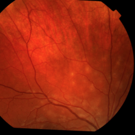
 Pericentral Retinitis Pigmentosa
Sep 6 2024 by Mauricio Bayram-Suverza, MD
A 65-year-old male patient reports experiencing bilateral blind spots that have gradually intensified over time. Genetic testing was unrevealing. The fundus autofluorescence image shows a hypoautofluorescent ring in the posterior pole, especially nasal to the nerve and along arcades.
Photographer: Mauricio Bayram-Suverza, Casey Eye Institute, OHSU.
Imaging device: Optos California
Condition/keywords: fundus autofluorescence (FAF), inherited retinal disease, nyctalopia, retinal dystrophy, retinitis pigmentosa
-
 Papilledema Probable to Pseudo Tumor Cerebri
Jun 30 2013 by Jason S. Calhoun
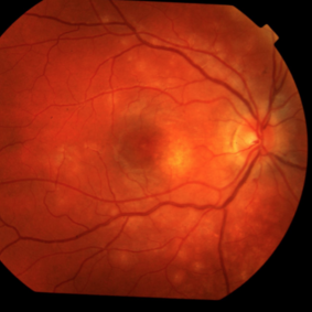
A 24-year-old female presented severe headaches and vision loss over the past 6-months. VA was 20/40 with pinhole 20/25, right eye and 20/25, left eye. Fundiscopic exam reveals 3-4 plus papilledema of both optic nerves. Patient has VF defects inferior, nasal-right eye and enlarged blind spot with an inferior arcuate defect present. Patient will get MRI and work-up with treatment in the hospital.
Photographer: Jason S. Calhoun, Mayo Clinic Jacksonville, Florida
Condition/keywords: papilledema
-
 Multiple Evanescent White Dot Syndrome (MEWDS)
Nov 1 2019 by Thomas A. Ciulla, MD, MBA, FASRS
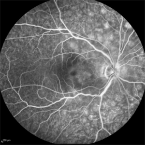
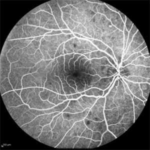
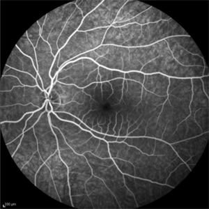
Fluorescein angiogram of an 18-year-old woman with multiple evanescent white dot syndrome (MEWDS). She noted a 5 day history of a new blind spot OD.
Condition/keywords: multiple evanescent white dot syndrome (MEWDS)
-
 AZOOR
AZOOR
Aug 24 2012 by Geoffrey G. Emerson, MD, PhD, FASRS
A 17-year-old healthy woman noticed a pacman-shaped scotoma in her temporal right vision. Acuity measured 20/20 and color vision measured 11/11. There was an enlarged physiologic blind spot on Humphry visual field testing. The fellow eye was normal.
Photographer: Geoffrey Emerson, MD, PhD, Retina Center, Minneapolis
Condition/keywords: scotoma
-
 Dye image
Apr 13 2024 by Monica King
Unknown diagnosis. 35yr old female. Blind spots started to form.
Photographer: Dr. Becker, Colorado Eye Institute
Condition/keywords: Unknown diagnosis
-
 Enlarged Blind Spot
Feb 13 2013 by From the Collections of Thomas M. Aaberg, MD and Thomas M. Aaberg Jr., MD
Peripillary guide resolved.
Condition/keywords: blind spot, peripapillary
-
 Multiple Evanescent White Dot Syndrome
Nov 1 2019 by Thomas A. Ciulla, MD, MBA, FASRS
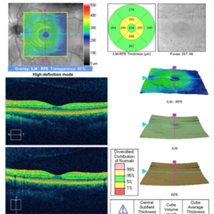
OCT OS of an 18-year-old woman with multiple evanescent white dot syndrome (MEWDS). She noted a 5 day history of a new blind spot OD.
Condition/keywords: multiple evanescent white dot syndrome (MEWDS)
-
 Multiple Evanescent White Dot Syndrome
Nov 1 2019 by Thomas A. Ciulla, MD, MBA, FASRS
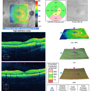
OCT OD of an 18-year-old woman with multiple evanescent white dot syndrome (MEWDS). She noted a 5 day history of a new blind spot OD.
Condition/keywords: multiple evanescent white dot syndrome (MEWDS)
-
 Multiple Evanescent White Dot Syndrome
Nov 1 2019 by Thomas A. Ciulla, MD, MBA, FASRS
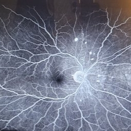
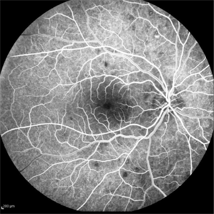
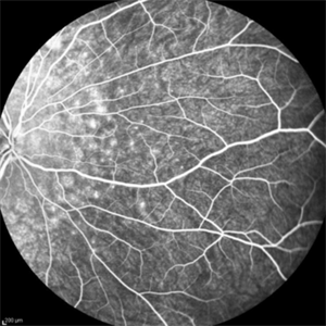
Fluorescein angiogram of an 18-year-old woman with multiple evanescent white dot syndrome (MEWDS). She noted a 5 day history of a new blind spot OD.
Condition/keywords: multiple evanescent white dot syndrome (MEWDS)
-
 Multiple Evanescent White Dot Syndrome
Nov 1 2019 by Thomas A. Ciulla, MD, MBA, FASRS
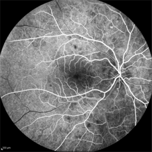
Fluorescein angiogram of an 18-year-old woman with multiple evanescent white dot syndrome (MEWDS). She noted a 5 day history of a new blind spot OD.
Condition/keywords: multiple evanescent white dot syndrome (MEWDS)
-
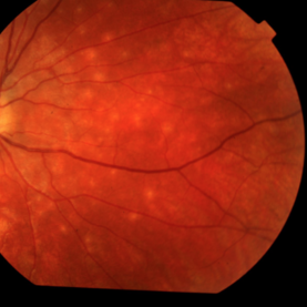
 Multiple Evanescent White Dot Syndrome (MEWDS)
Nov 1 2019 by Thomas A. Ciulla, MD, MBA, FASRS
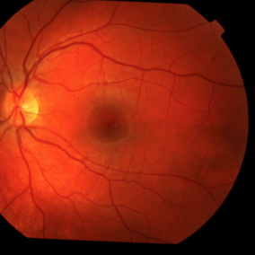
Fundus photograph of an 18-year-old woman with multiple evanescent white dot syndrome (MEWDS). She noted a 5 day history of a new blind spot OD.
Condition/keywords: multiple evanescent white dot syndrome (MEWDS)
-
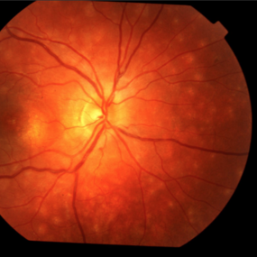
 Multiple Evanescent White Dot Syndrome (MEWDS)
Nov 1 2019 by Thomas A. Ciulla, MD, MBA, FASRS
Fundus photograph of an 18-year-old woman with multiple evanescent white dot syndrome (MEWDS). She noted a 5 day history of a new blind spot OD.
Condition/keywords: multiple evanescent white dot syndrome (MEWDS)
-
 Multiple Evanescent White Dot Syndrome (MEWDS)
Nov 1 2019 by Thomas A. Ciulla, MD, MBA, FASRS
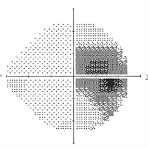
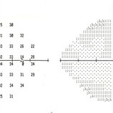
Visual field of an 18-year-old woman with multiple evanescent white dot syndrome (MEWDS). She noted a 5 day history of a new blind spot OD.
Condition/keywords: multiple evanescent white dot syndrome (MEWDS)
-
 Multiple Evanescent White Dot Syndrome (MEWDS)
Nov 1 2019 by Thomas A. Ciulla, MD, MBA, FASRS
Fluorescein angiogram of an 18-year-old woman with multiple evanescent white dot syndrome (MEWDS). She noted a 5 day history of a new blind spot OD.
Condition/keywords: multiple evanescent white dot syndrome (MEWDS)
-
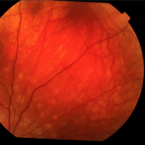
 New Retinal Detachment 6w s/p RD repair
Nov 16 2023 by Virginia Gebhart
13 year old male presented with new blind spot 6 weeks s/p RD repair with cryo/scleral buckle/prophylaxis laser with gas bubble. New RD involving the macula, posterior to scleral buckle, secondary to PVD. Small gas bubble remaining. Pt was brought back to OR for repeat PPV and silicone oil repair
Photographer: Virginia Gebhart
Imaging device: Optos
Condition/keywords: gas bubble, Retinal Detachment, retinal detachment of the macula, scleral buckle
-
 Optic Disc Drusen
Mar 27 2013 by Henry J. Kaplan, MD
Perimetry demonstrates slightly enlarged blind spot in the same patient #5.
Condition/keywords: drusen of optic disc, optic disc drusen, optic nerve drusen
-
---thumb.JPG/image-square;max$300,300.ImageHandler) Papilledema Probable to Pseudo Tumor Cerebri
Papilledema Probable to Pseudo Tumor Cerebri
Jun 30 2013 by Jason S. Calhoun
A 24-year-old female presented severe headaches and vision loss over the past 6 months. VA was 20/40 with pinhole 20/25, right eye and 20/25, left eye. Fundiscopic exam reveals 3-4 plus papilledema of both optic nerves. Patient has VF defects inferior, nasal-right eye and enlarged blind spot with an inferior arcuate defect present. Patient will get MRI and work-up with treatment in the hospital.
Photographer: Jason S. Calhoun, Mayo Clinic Jacksonville, Florida
Condition/keywords: papilledema
-
---thumb.JPG/image-square;max$300,300.ImageHandler) Papilledema Probable to Pseudo Tumor Cerebri
Papilledema Probable to Pseudo Tumor Cerebri
Jun 30 2013 by Jason S. Calhoun
A 24-year-old female presented severe headaches and vision loss over the past 6 months. VA was 20/40 with pinhole 20/25, right eye and 20/25, left eye. Fundiscopic exam reveals 3-4 plus papilledema of both optic nerves. Patient has VF defects inferior, nasal right eye and enlarged blind spot with an inferior arcuate defect present. Patient will get MRI and work-up with treatment in the hospital.
Photographer: Jason S. Calhoun, Mayo Clinic Jacksonville, Florida
Condition/keywords: papilledema
 Loading…