Search results (5 results)
-
 Dislocated Crystalline Lens
Mar 19 2024 by Annaka Gooding
Ultra Wide field fundus photography of a 70 year old male who presented to clinic with a sudden increase of vision due to dropped crystalline lens secondary to severely dense cataract. Patient reported seeing a full black circle in his inferior visual field. Patient's visual acuity at time of visit was 20/100 with a +5.00 diopter lens. The physician recommended surgical intervention, and discussed surgery for PPV/PPL/IOL implantation with an ACIOL.
Photographer: Annaka Gooding, CPO
Imaging device: Optos California RGB
Condition/keywords: dislocated crystalline lens, fundus photography, inferior retina, OPTOS CALIFORNIA RGB, Right Eye, Ultra-wide field retinal imaging
-
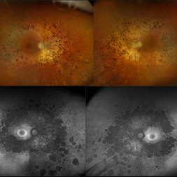 Retinitis Pigmentosa
Nov 7 2023 by Jolee Rodriguez
Bilateral fundus photography and fundus autofluorescence imaging of a 62-year-old male with Retinitis Pigmentosa. Patient reported visual field defects and dark adapting issues. Patient's vision at the time images were taken were sc20/20 of the right eye and sc20/25 of the left eye. Dr. Sutherland determined that based on the patient's lack of family history, the most likely route of inheritance is autosomal recessive.
Photographer: Jolee Rodriguez
Imaging device: Optos California RGB
Condition/keywords: autofluorescence imaging, fundus photography, hereditary retinal dystrophy, Optos, OPTOS CALIFORNIA RGB, retinitis pigmentosa, ultra-wide field imaging, Ultra-wide field retinal imaging, ultra-widefield image
-
 Eales Disease
Jan 31 2025 by Thirumalesh Mochi Basavaraj, MD
Ultra-wide field image of a 24 year old young healthy adult male with a visible sea fan neovascularization with partial PVD with vitreous and subhyaloid hemorrhage.
Photographer: Puttaswamy
Condition/keywords: Eales disease, sea fan, Ultra-wide field retinal imaging
-
 Ocular Ischemic Syndrome
Jun 27 2023 by Mauricio Bayram-Suverza, MD
Fundus photograph of a 72-year-old man referred to the retina department due to long-term, painless vision loss in his left eye. Following fluorescein angiography, prolonged arteriovenous transit time and hypoperfusion were detected. Consequently, the patient was advised to undergo cardiological evaluation.
Photographer: Mauricio Bayram-Suverza, Fundación Hospital Nuestra Señora de la Luz
Imaging device: Optos California
Condition/keywords: Fluorescein angiography, ocular ischemic syndrome, Ultra-wide field retinal imaging
-
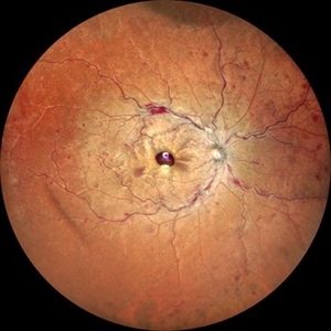 Ultra-Wide Field Image of Central Retinal Vein Occlusion with Foveal Hemorrhage
Apr 17 2025 by Malvika Singh
Ultra- wide field fundus photograph of a 41 year-old male, with a central retinal vein occlusion and a foveal sub-internal limiting membrane hemorrhage.
Photographer: Dr Malvika Singh, Retina Foundation, Ahmedabad, India
Imaging device: Mirante SLO/OCT
Condition/keywords: central retinal vein occlusion (CRVO), macular hemorrhage, Ultra-wide field retinal imaging
 Loading…