Search results (25 results)
-
 Macroaneurysm
Jul 11 2016 by Manish Nagpal, MD, FRCS (UK), FASRS
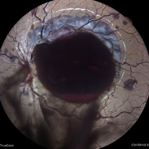
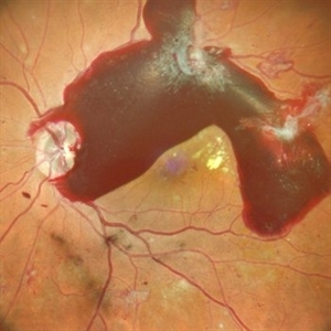
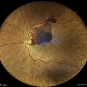
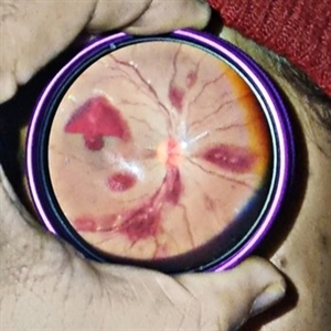
Fundus photography of a 45 year old male who presented with a macroaneurysm in his right eye with a spill over de haemoglobinised sub hyaloid haemorrhage
Photographer: Pooja Barot
Condition/keywords: macroaneurysm, subhyaloid hemorrhage
-
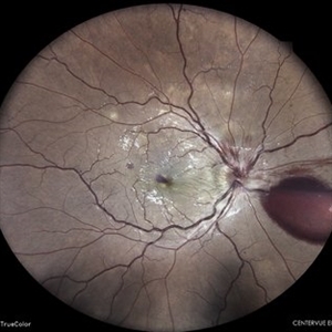
 Subhyaloid Haemorrhage
Feb 5 2023 by RAKESH SHAH, MS DNB FACS FRF FICO MBA
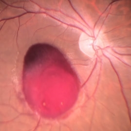
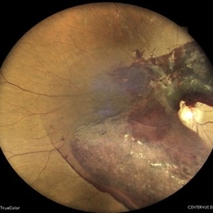
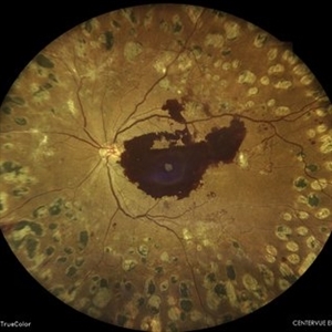
YOUNG MALE 24 YEARS COMES WITH SPONTANEOUS LOSS OF VISION AFTER HEAVY EXERCISES IN GYM
Photographer: DR RAKESH SHAH
Condition/keywords: Sub hyaloid haemorrhage
-
 VALSALVA RETINOPATHY
Jun 6 2023 by Akansha Sharma
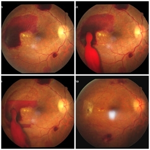
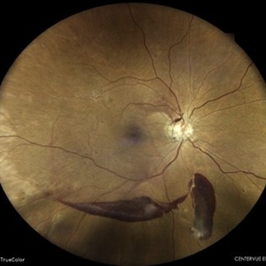
COLOUR FUNDUS PHOTOGRAPH OF A 21 YEAR OLD MALE WITH VALSALVA RETINOPATHY
Photographer: Dr. Urmil Shah, Dr. Denish Patel, Dr. Akansha Sharma, Bharati Eye Clinic, Ahmedabad, Gujarat
Condition/keywords: Sub hyaloid haemorrhage, SUB RETINAL HEMORRHAGE, valsalva retinopathy
-
 10 days after Green LASER hyaloidotomy
Jun 29 2022 by Tandava Krishnan
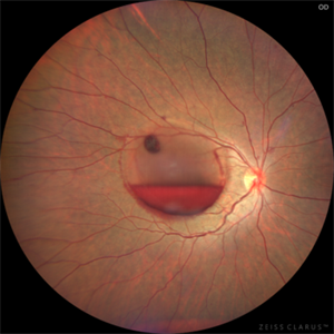
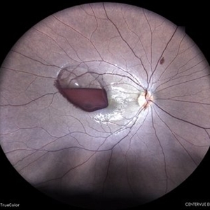
This is the left eye fundus picture of a 35 year old gentleman 10 days after LASER hyaloidotomy
Photographer: Dr Tandava Krishnan, Dr Agarwal's eye Hospital , Hyderabad, India
Condition/keywords: Green LASER, Sub hyaloid haemorrhage
-
 Hyaloid Butterfly
Mar 13 2025 by Gustavo Uriel Fonseca Aguirre
Axial ultrasound showing a phakic eye with vitreous hemorrhage, hyaloids impregnated with blood, hyalochisis (butterfly-shaped), subhyaloid hemorrhage, and retinal tractions involving the macular area.
Photographer: Gustavo U. Fonseca Aguirre, Hospital Conde de Valenciana, Ciudad de México
Condition/keywords: Hyaloschisis, Sub hyaloid haemorrhage, Vitreous hemorrhage
-
 Pre retinal Hemorrhage
Jul 5 2024 by Anjana Mirajkar, MS Ophthalmology
An intra operative image of a pre retinal hemorrhage like a sub hyaloid hemorrhage at the macular area.
Photographer: Dr. Anjana Mirajkar -Retina Foundation, Ahmedabad
Condition/keywords: Sub hyaloid haemorrhage
-
 Proliferative Diabetic Retinopathy
May 24 2024 by Anjana Mirajkar, MS Ophthalmology
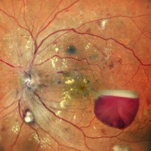
A central photo of a 50 year old male case of PDR showing a sub-hyaloid hemorrhage with cotton wool spots , hard exudates at the fovea with dot and blot hemorrhages.
Photographer: Dr. Anjana Mirajkar -Retina Foundation, Ahmedabad
Imaging device: Mirante-Nidek
Condition/keywords: proliferative diabetic retinopathy (PDR), Sub hyaloid haemorrhage
-
 PROLIFERATIVE DIABETIC RETINOPATHY
May 31 2023 by Akansha Sharma
colour fundus photograph of a 56 year old male with fibrovascular proliferations and subhyaloid hemorrhage in a case of proliferative diabetic retinopathy
Photographer: Dr. Urmil Shah, Dr. Akansha Sharma, Dr. Denish Patel
Condition/keywords: PDR, proliferative diabetic retinopathy (PDR), Sub hyaloid haemorrhage
-
 SUB HYALOD HEMORRHAGE
Oct 18 2023 by ANKIT JAIN
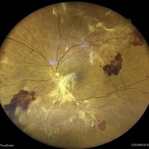
WIDEFIELD FUNDUS IMAGE OF RIGHT EYE OF 53 YEAR OLD MALE, KNOWN CASE OF DIABETES MELLITUS TYPE 2 FROM 3 YEARS WITH SUB-HYALOID HEMORRGAE IN CASE OF PROLIFERATIVE DIABETIC RETINOPATHY
Photographer: DR ANKIT JAIN
Imaging device: MIRANTE
Condition/keywords: diabetes, proliferative diabetic retinopathy (PDR), Sub hyaloid Haemorrhage
-
 Sub hyaloid haemorrhage 10 minutes after LASER hyaloidotomy
Jun 29 2022 by Tandava Krishnan
This is the left eye fundus picture of a 35 year old man, 10 minutes after Green LASER Hyaloidotomy showing clearing of blood from the sub hyaloid space into the vitreous
Photographer: Dr Tandava Krishnan, Dr Agarwal's eye Hospital, Hyderabad, India
Condition/keywords: Green LASER, Sub hyaloid Haemorrhage
-
 Sub Hyaloid Haemorrhage after Valsalva manoeuvre
Jun 29 2022 by Tandava Krishnan
Left eye fundus picture of a 35 year old gentleman showing sub hyaloid haemorrhage after Valsalva manoeuvre
Photographer: Dr Tandava Krishnan, Dr Agarwal's Eye Hospital, Hyderabad, India
Condition/keywords: Green LASER, Sub hyaloid haemorrhage
-
 Sub Hyaloid Haemorrhage not involving fovea
Feb 3 2020 by Sham Talati, DOMS
A diabetic case of subhyaloid hemorrrhage not involving fovea.
Photographer: Dr. Sham Talati,Retina Foundation,Ahmedabad
Imaging device: Nidek Mirante
Condition/keywords: proliferative diabetic retinopathy (PDR), subhyaloid hemorrhage
-
 Sub Hyaloid Hemorrhage
Jan 29 2025 by Vishal Agrawal, MD, FRCS,FACS,FASRS
A 45-year-old male patient presented with decreased vision in Left Eye. On fundus examination, a boat-shaped sub hyaloid hemorrhage was noted. YAG hyaloidotomy was performed and the patient recovered with a vision of 20/20.
Photographer: Dr Ayushi Gupta
Imaging device: Clarus 700
Condition/keywords: Sub hyaloid haemorrhage, YAG HYALOIDOTOMY
-
 SUB HYALOID HEMORRHAGE
Feb 28 2023 by Akansha Sharma
COLOUR FUNDUS PHOTOGRAPH OF A 55 YEAR OLD FEMALE WITH SUB HYALOID HEMORRHAGE
Photographer: Dr. Urmil Shah, Dr. Denish Patel, Dr. Akansha Sharma, Bharati Eye Hospital, Ahmedabad, Gujarat
Condition/keywords: SHH, Sub hyaloid haemorrhage
-
 Sub Hyaloid Hemorrhage
Dec 14 2024 by Harshit Krishna
Fundus photography in a 30 year old leukemic female, showing sub hyaloid hemorrhage drained with YAG hyaloidotomy.
Photographer: Dr. Harshit Krishna Malavat
Condition/keywords: Sub hyaloid haemorrhage
-
 SUB HYLOID HAEMORRHAGE IN A CASE OF RETINAL MACROANEURYSM
Jun 9 2022 by Nivesh Gupta
Fundus photograph of a 34 year old female with sub hyaloid haemorrhage in a case of retinal macroaneurysm.
Photographer: DR. NIVESH GUPTA, RETINA FELLOW , RETINA FOUNDATION, AHMEDABAD
Imaging device: NIDEK MIRANTE
Condition/keywords: macroaneurysm, sub internal limiting membrane haemorrhage
-
 Subhyaloid Hemorrhage
Mar 20 2024 by Othman Karmane
Fundus photograph of a 67 year-old man, with a spontaneous subhyaloid hemorrhage.
Photographer: Karmane Othman
Condition/keywords: Sub hyaloid Haemorrhage
-
 Subhyaloid Hemorrhage
Jul 31 2024 by Arthi Mohankumar , MS,MRCS ED, FICO,FAICO
A 35 year old male presented with complaints of seeing a black spot in left eye for past one day after working out in the gym the previous day. He has history of uncontrolled diabetes and hypertension. Fundus exam of the left eye revealed a sub hyaloid hemorrhage nasal to the disc with minimal background Diabetic and hypertensive changes. His baseline CBG was 200 mg/dl and BP was 170/100 He was suggested observation initially considering the nasal location. But patient found the scotoma very disturbing and eventually underwent yag hyaloidotomy
Photographer: Arthi Mohankumar
Condition/keywords: Sub hyaloid haemorrhage, valsalva retinopathy
-
 Subhyaloid Hemorrhage
Mar 1 2025 by Vishal Agrawal, MD, FRCS,FACS,FASRS
A 37-year-old male presented with sudden diminution of vision in the right eye. On fundus examination boat shaped sub hyaloid hemorrhage was noted and a YAG hyaloidotomy was performed.
Photographer: Dr Ayushi Gupta
Imaging device: Clarus 700
Condition/keywords: Sub hyaloid haemorrhage, YAG HYALOIDOTOMY
-
 Subhyaloid Hemorrhage From a Macroaneurysm
Jan 30 2024 by Akansha Sharma
Color fundus photograph of a 58 year old hypertensive and diabetic female patient with lasered proliferative diabetic retinopathy developing a subhyaloid hemorrhage from a macroaneurysm.
Photographer: Dr. Akansha Sharma, Bharati Eye Hospital
Condition/keywords: florid type PDR, macroaneurysm, proliferative diabetic retinopathy (PDR), SHH, Sub hyaloid haemorrhage
-
 Subhyaloid Hemorrhage in a Case of Lasered Proliferative Diabetic Retinopathy
Jun 24 2024 by Akansha Sharma
Color fundus photograph of a 47 year old female with subhyaloid hemorrhage in a case of lasered proliferative diabetic retinopathy.
Photographer: Dr. Akansha Sharma, Bharati Eye Hospital
Condition/keywords: PDR, proliferative diabetic retinopathy (PDR), SHH, Sub hyaloid haemorrhage
-
 Subhyaloid Hemorrhage in a Case of Proliferative Diabetic Retinopathy
Mar 26 2024 by Akansha Sharma
Color fundus photograph of a 56 year old male patient with subhyaloid hemorrhage in a case of proliferative diabetic retinopathy.
Photographer: Dr. Akansha Sharma, Bharati Eye Hospital
Condition/keywords: PDR, proliferative diabetic retinopathy (PDR), SHH, Sub hyaloid haemorrhage
-
 SUBHYALOID HEMORRHAGE IN A CASE OF VALSALVA RETINOPATHY
May 31 2023 by Akansha Sharma
COLOUR FUNDUS PHOTOGRAPH OF A 20 YEAR OLD MALE WITH SUBHYALOID HEMORRHAGE IN A CASE OF VALSALVA RETINOPATHY
Photographer: Dr. Urmil Shah, Dr. Akansha Sharma, Dr. Denish Patel
Condition/keywords: Sub hyaloid Haemorrhage, valsalva retinopathy
-
 Thrombocytopenia
Sep 24 2024 by DR Rohit Gupta
Fundus photography of a 16 year old female suffering from severe thrombocytopenia. On fundus examination, multiple roth spots and subhyaloid hemorrhage were seen.
Photographer: Dr Rohit gupta
Imaging device: Samsung S21
Condition/keywords: ANEMIC RETINOPATHY, hemorrhage, leukemia, retinal hemorrhage, Roth spots, Sub hyaloid haemorrhage, thrombocytopenia
-
 Venous Stasis Retinopathy
Jun 27 2024 by Akansha Sharma
Color fundus photograph of a 31 year old male patient with venous stasis retinopathy.
Photographer: Dr. Akansha Sharma, Bharati Eye Hospital
Condition/keywords: cystoid macular edema (CME), Disc Edema, SHH, Sub hyaloid haemorrhage
 Loading…