Search results (11 results)
-
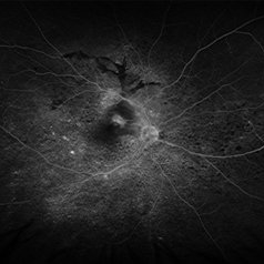
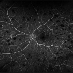 Roth Spots
Oct 26 2022 by Denica Rodriguez
Roth spots during optos FA on a 68 year old female with retinal hemorrhage effecting her left eye. Patient was referred for non-proliferative diabetic retinopathy without macular edema.
Photographer: Denica Rodriguez & Zachary Seim
Imaging device: Optos California
Condition/keywords: Diabetes, FLUORESCEIN ANGIOGRAPHY, left eye, Optos, Retina, Roth Spots, ultra-wide field imaging
-
 Venous Beading
Apr 30 2021 by Shivani Reddy, MD
This is a fluorescein angiogram image capturing a beautiful example of different stages of venous beading in diabetic retinopathy all in one frame. This patient also has various microangiopathic findings including microaneurysms, venous loops and capillary dropout. This patient is a 41 y/o male with a history of type 1 diabetes, presenting for his first eye exam in years.
Imaging device: Optos FA
Condition/keywords: capillary dropouts, nonproliferative diabetic retinopathy, proliferative diabetic retinopathy (PDR), retinal ischemia, venous beading
-
 Acute Central Retinal Artery Occlusion
Jul 27 2022 by Becca Harris
Ultra widefield FA/ICG of a 24 year old female with an acute central retinal artery occlusion affecting the right eye. Patient presented with extreme headaches following DAVF surgery the previous day. Patient has Factor VIII deficiency and had a cerebral venous thrombosis 9 years ago and lost vision in the right eye at that time. Patient has history of optic sheath fenestration OU and craniotomy. On initial evaluation, she had a CRAO as well as diffuse choroidal nonperfusion noted on optos FA. Suspect nonperfusion to third and sixth nerve leading to palsy. Occlusion of vasculature in the setting of recent endovascular embolization of fistulas in the CNS. Discussed diagnosis and poor prognosis with parents and patient. Patient had no light perception at the time of her initial appointment.
Photographer: Becca Harris
Imaging device: Optos California
Condition/keywords: Choroidal non-perfusion, fluorescein angiogram (FA), indocyanine green (ICG) angiography, non-perfusion, Optos, Right Eye, ultra-wide field imaging
-
 Discrete Choroidal Hemangioma
Jan 7 2018 by John S. King, MD
49-year-old WF seen for annual, routine exam, and sent here because could not refract to better than 20/40. Optos Photos and FA showed minimal to no findings, while topcon photos showed a well curcumscribed red/orange lesion with high internal reflectivity on Bscan, and sparing of choriocapillaris on OCT; no SRF, exudates, or other findings present.
Imaging device: Optos FA
Condition/keywords: choroidal hemangioma
-
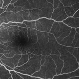
 Ischemic HRVO with Macular Edema
Mar 7 2024 by Jenn Geelan
Optos FA of an 80 year old female.
Photographer: Jenn Geelan
Imaging device: Optos California
Condition/keywords: FA late phase, hemicentral retinal vein occlusion, ischemic CRVO, macular edema
-
 Myope With Staphyloma and Vitreous Detachment
Jun 11 2016 by Philip J. Polkinghorne, MD
Fundus autofluorescence of a myope with PVD and staphyloma.
Imaging device: Optos FAF
Condition/keywords: degenerative myopia, myopia, staphyloma
-
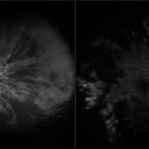
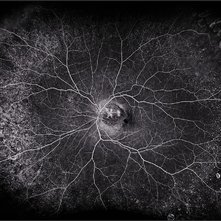
 Optos FA of Harada's Disease
Jun 22 2013 by James A Eadie, MD
Optos fluorescein angiogram of a 22-year-old woman with Harada's disease. An exudative detachment is billowing in the foreground at the bottom of the image.
Photographer: John Peterson
Imaging device: Optos camera
Condition/keywords: Harada's disease, Vogt-Koyanagi-Harada
-
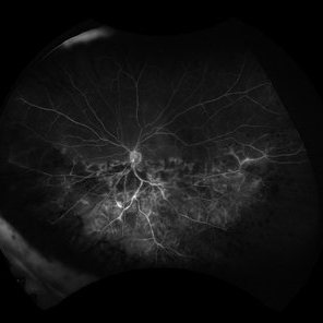
 Panuveitis
Jul 12 2024 by Korey Starkey
Ultra widefield Optos FA of 59 year old female presents with panuveitis in both eyes. Patients vision was VA OS: Dcc20/60-2 at time of visit.
Photographer: Korey Starkey
Imaging device: Optos
Condition/keywords: FLUORESCEIN ANGIOGRAPHY, hyperfluorescence, Optos, Panuveitis, ultra-wide field imaging, Uveitis
-
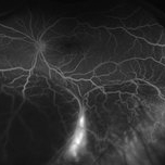
 PPCNVM and Peripheral Drusen Seen on Optos FA
Apr 22 2020 by John S. King, MD
72-year-old white male c/o of distortion OS for about 2 months. 20/100 OS, normotensive, small grey-green subretinal area just temporal to the optic disc. FA shows leakage c/w a ppcnvm; there is some SR and IR leakage as well as staining of peripheral drusen and some window defects from cobblestone. Avastin was adminstered.
Photographer: Asli Ahmed
Imaging device: CA
Condition/keywords: drusen, peripapillary choroidal neovascularization (PPCNVM)
-
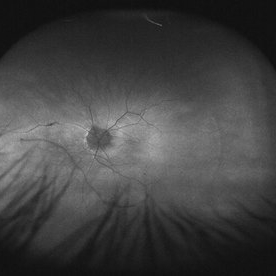
 Uveal Effusion Syndrome
Jan 7 2025 by Drew Mitchell
Optos FA Late of Uveal Effusion Syndrome
Photographer: Drew Mitchell, OCT-C
Imaging device: Optos California
Condition/keywords: FA late phase, Optos, uveal effusion
-
 VKH Pseudotumor – Fluorescein Angiography
May 11 2025 by Felipe Murati
Fluorescein angiography image from a 36-year-old woman with chronic Vogt-Koyanagi-Harada (VKH) syndrome showing a pseudotumor-like lesion with late-phase staining and no active leakage. The image highlights subretinal fibrosis in the right eye, stable under long-term immunosuppressive therapy with mycophenolate mofetil and adalimumab. No signs of active choroiditis are present, confirming a quiescent phase.
Photographer: Felipe A. Murati, MD, University of Arizona
Imaging device: Optos California, fluorescein angiography modality
Condition/keywords: choroiditis, Fluorescein angiography, granulomatous uveitis, Optos FA, pseudotumor, subretinal fibrosis, VKH, Vogt-Koyanagi-Harada
 Loading…