Search results (13 results)
-
 Ectopia Lentis
Jan 21 2021 by Jamin S. Brown, MD
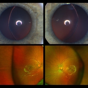
This image serial demonstrates a patient with simple ectopia lentis. Anterior segment photographs in the upper panel demonstrate nasally subluxated crystalline lenses. Widefield fundus photography shows a "pseudo-buckle" which is the result of an optical effect due to the lens subluxation (artifactual image enlargement). Also note the juvenile macular reflex in this young patient. Ectopia lentis can present isolated ("simple") or in combination with various systemic defects (Marfan's syndrome, Weil-Marchesani syndrome or Ehlers-Danlos syndrome to name a few). Isolated ectopia lentis can be hereditary and causative genes have been identified as ADAMTSL4 located on chromosome 4 and FBN1 gene located on chromosome 15. Defects in the genes cause weakness in the zonular fibers which can lead to lens dislocation. Lastly, various ocular disorders such as Aniridia, Axenfeld-Rieger, Pseudoexfoliation or Trauma may also result in lens dislocation or subluxation.
Photographer: Stefanie Palmer CRA, Retina Vitreous Surgeons of CNY
Condition/keywords: dislocated lens, ectopia lentis
-
 Dislocation of the Crystalline Lens with a Retinal Detachment
Apr 21 2025 by Hrishikesh Naik, MS
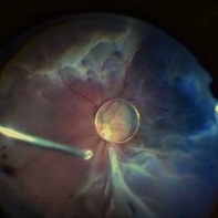
An intraoperative screen grab shows a dislocation of the crystalline lens along with an associated rhegmatogenous retinal detachment in a case of Marfan’s syndrome. The case was managed by a combined PPV-SB procedure. A vitrectomy cutter is seen at the left.
Photographer: Hrishikesh Naik
Condition/keywords: intraoperative, lens dislocation, Marfan's syndrome, Retinal Detachment, vitrectomy
-
 Bullous RD With Dislocated Lens
Apr 3 2018 by Navneet Mehrotra, DNB
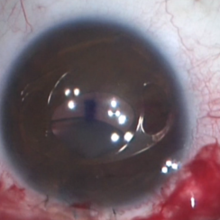
Dislocated clear lens and associated retinal detachment in a young patient with Marfan's syndrome.
Photographer: Navneet Mehrotra
Imaging device: Sony 3 chip camera
Condition/keywords: dislocated crystalline lens, Marfan's syndrome
-
 Artisan Lens
Aug 23 2018 by Jessica G Lee, MD
Intraoperative photo of a 13 -year-old boy with Marfan's syndrome with dislocated crystalline lens, undergoing procedure for placement of Artisan lens.
Condition/keywords: intraocular lens (IOL), Marfan's syndrome
-
---thumb.jpg/image-square;max$300,300.ImageHandler) Flexibility in Marfan's Syndrome
Flexibility in Marfan's Syndrome
Dec 19 2012 by Eric A. Postel, MD
Color photo demonstrating flexibility and long fingers in a patient with Marfan's Syndrome
Condition/keywords: Marfan's syndrome
-
 Hands of 3 generations of a Marfan's family
Dec 19 2012 by Eric A. Postel, MD
Color photograph of the hands of patients from 3 generations of a family with Marfan's Syndrome.
Condition/keywords: Marfan's syndrome
-
Iridodonesis
Feb 8 2016 by Andrea Arriola-Lopez, MD MSc
Marfan syndrome patient, subluxated lens and iridodonesis. BCVA 20/60.
Photographer: Andrea Elizabeth Arriola-Lopez MD MSc
Condition/keywords: iris, Marfan's syndrome, retina, subluxation of lens
-
 Marfan's Syndrome Ocular Manifestation
Jan 30 2015 by H. Michael Lambert, MD
Subluxed lens and zonular dehiscence.
Condition/keywords: Marfan's syndrome, subluxation of lens
-
 Marfan's Syndrome Ocular Manifestation
Jan 30 2015 by H. Michael Lambert, MD
Subluxed lens and zonular dehiscence.
Condition/keywords: Marfan's syndrome, subluxation of lens
-
 Marfan's Syndrome Ocular Manifestation
Jan 30 2015 by H. Michael Lambert, MD
Subluxed lens and zonular dehiscence.
Condition/keywords: Marfan's syndrome, subluxation of lens
-
 Marfan's Syndrome Ocular Manifestation
Jan 30 2015 by H. Michael Lambert, MD
Subluxed lens and zonular dehiscence.
Condition/keywords: Marfan's syndrome, subluxation of lens
-
 Myopia with Lattice Degeneration and White Without Pressure in the Setting of Marfan's Syndrome
Aug 31 2020 by Sophia El Hamichi, MD
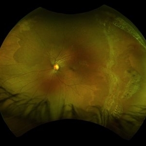
A 1-year-old female with Marfan's syndrome, myopia OU, congenital nystagmus and exotopia OD. Ultra-wide field imaging of her left eye showed lattice degeneration with atrophic retinal holes temporally, in addition to multiple sections of white without pressure.
Imaging device: Optos
Condition/keywords: atrophic retinal hole, lattice degeneration, Marfan's syndrome, myopia, Optos, ultra-wide field imaging
-
 Vitreous Wrap Around The Dislocated Nucleus
Nov 14 2019 by Deepak Bhojwani, MS
Intraoperative Image of a spontaneous dislocation of lens in a Marfanoid patient.
Photographer: Dr Deepak Bhojwani
Imaging device: Sony External Video Recording Camera
Condition/keywords: dislocated crystalline lens, Marfan's syndrome
 Loading…