Search results (18 results)
-
 Acute Posterior Multifocal Placoid Pigment Epitheliopathy
Feb 20 2024 by Soobien Lee
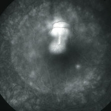
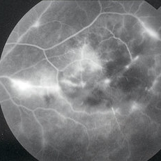
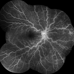
Fluorescein angiogram of a 20-year-old caucasian female with viral prodrome and vision loss OS>OD secondary to Acute Posterior Multifocal Placoid Pigment Epitheliopathy (APPME). Early blockage with late hyperfluorescent leakage can be seen on fluorescein angiography of the left eye.
Photographer: Ashley Metzger, Elman Retina Group
Imaging device: Optos Ultra-Widefield Fluorescein Angiography
Condition/keywords: acute posterior multifocal placoid pigment epitheliopathy (APMPPE), bacilliary layer detachment, FA, FA late phase, FA late phase leakage, fluorescein angiogram (FA), Optos, uveitis, white dot syndrome
-
 ROP FA OD
Apr 27 2018 by Brenda Fallas
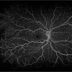
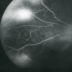
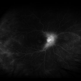
4-month-old baby with regressed ROP post-Avastin.
Photographer: Brenda Fallas, Bascom Palmer Eye Institute, Miami, FL
Imaging device: RETCAM III 130 degree lens mongtage
Condition/keywords: FA late phase leakage, fluorescein angiogram (FA), retina, retinopathy of prematurity (ROP)
-
 ROP FA OS
Apr 27 2018 by Brenda Fallas
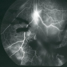
4-month-old baby with regressed ROP post-Avastin.
Photographer: Brenda Fallas, Bascom Palmer Eye Institute, Miami, FL
Imaging device: RETCAM III 130 degree lens montage
Condition/keywords: FA late phase leakage, fluorescein angiogram (FA), retinopathy of prematurity (ROP)
-
 Angiographic Storm: Fluorescein Leakage in Retinal Vasculitis
Nov 17 2025 by SHRADDHA RAJ SHRIVASTAVA
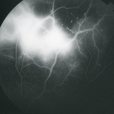
This left eye montage fundus fluorescein angiography (FFA) image of a 19 year old male with idiopathic retinal vasculitis, having skip vasculitic lesions predominantly involving retinal veins. There are areas of blocked fluorescence due to intraretinal hemorrhages, the involved veins have filling defects and occlusions, leading to formation of numerous collateral channels. The inflamed vessels also show perivascular fuzzy hyperfluorescent stain due to leakage of dye. We can also see multiple peripheral capillary non perfusion (CNP) areas, with a 'hot disc', suggestive of ongoing inflammation.
Photographer: Dr. Shraddha Raj Shrivastava
Imaging device: Nidek Mirante SLO/OCT (Confocal scanning/Spectral domain OCT)
Condition/keywords: FA late phase leakage, Fundus Fluorescein Angiography, idiopathic retinal vasculitis, optic disc leakage, VASCULITIS
-
 Behcet's Eye Disease
Apr 10 2017 by Deepak Bhojwani, MS
A 19-year-old boy presented with with recurrent oral and genital ulcers along with blurring of vision. Systemically he was HLA B-51 Positive suggesting Behcet's Disease. The FFA photograph depicts the classic small vessel immune mediated vasculitis predominantly affecting the capillaries (capillaropathy).
Photographer: DEEPAK BHOJWANI, RAGHUDEEP EYE HOSPITAL, AHMEDABAD.
Condition/keywords: Behcet's Disease, Behcet's uveitis, FA late phase leakage
-
 Bilateral Central Serous Retinopathy
Mar 26 2019 by Gary R. Cook, MD, FACS
Late-phase frame of FA of 37-year-old white male with acute CSR OD showing pooling of dye beneath the small central RPED centrally, a smokestack-type leak from the RPE defect just above it, and mild late pooling of dye outlining the large neurosensory macular detachment; VA = 20/80-1.
Imaging device: Topcon VT-50
Condition/keywords: central serous retinopathy (CSR), FA late phase, FA late phase leakage, neurosensory detachment of retina
-
 Central Retinal Vein Occlusion With Macular Edema
Aug 29 2018 by Olivia Rainey
Ultra-widefield fluorescein angiogram of an 58-year-old male with a central retinal vein occlusion with macular edema affecting his right eye. Fluorescein showed delayed transit with late leakage.
Photographer: Olivia Rainey
Imaging device: Optos
Condition/keywords: central retinal vein occlusion (CRVO), FA late phase leakage, fluorescein angiogram (FA), Optos, tortuous vessels, ultra-wide field imaging
-
 Central Serous Retinopathy
Mar 26 2019 by Gary R. Cook, MD, FACS
32-year-old white female with acute CSR OS; 10-minute late frame of FA OS showing diffusion of dye from earlier pinpoint leak in inferonasal macula; VA = 20/30-2.
Imaging device: Topcon VT-50
Condition/keywords: central serous retinopathy (CSR), FA late phase leakage, fluorescein angiogram (FA)
-
 CME
CME
May 3 2013 by Suber S. Huang, MD, MBA, FASRS
CME.
Imaging device: Retina Diseases Imaging Analysis Reading Center
Condition/keywords: angiographic macular leakage, cystoid macular edema (CME), FA late phase leakage
-
 Eales Disease
Apr 1 2019 by Gary R. Cook, MD, FACS
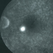
Late-phase fluorescein angiogram image of the right eye of a 23-year-old Vietnamese female with Eales disease showing vascular leakage and staining in the posterior pole OD.
Imaging device: Topcon VT-50
Condition/keywords: Eales disease, FA late phase, FA late phase leakage, fluorescein angiogram (FA), retinal vasculitis
-
 Eales Disease
Apr 1 2019 by Gary R. Cook, MD, FACS
Mid-peripheral retinal vascular changes on late-phase fluorescein angiography of a 23-year-old Vietnamese female with Eales disease.
Imaging device: Topcon VT-50
Condition/keywords: Eales disease, FA late phase, FA late phase leakage, fluorescein angiogram (FA), retinal vasculitis
-
 Eales Disease
Apr 1 2019 by Gary R. Cook, MD, FACS
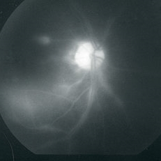
Late-phase (5 minutes) fluorescein angiogram image of the nasal mid-periphery of the left eye of a 23-year-old Vietnamese female with Eales Disease showing multiple areas of NVE and some disc leakage.
Imaging device: Topcon VT-50
Condition/keywords: Eales disease, FA late phase, FA late phase leakage, fluorescein angiogram (FA), neovascularization elsewhere (NVE)
-
 Eales Disease
Apr 1 2019 by Gary R. Cook, MD, FACS
Mid-phase (70 seconds) fluorescein angiogram image of the inferior periphery OS of a 20-year-old Vietnamese male with Eales Disease; there is bright hyperfluorescence from a focus of NVE below the optic disc and blocked fluorescence from vitreous hemorrhage in the eye.
Imaging device: Topcon VT-50
Condition/keywords: Eales disease, FA late phase, FA late phase leakage, fluorescein angiogram (FA), neovascularization elsewhere (NVE), vitreous hemorrhage
-
 Eales Disease
Apr 1 2019 by Gary R. Cook, MD, FACS
Late-phase (5 minutes) fluorescein angiogram image of a 20-year-old Vietnamese male with Eales Disease showing retinal vascular changes and intense leakage from peripheral NVE.
Imaging device: Topcon VT-50
Condition/keywords: Eales disease, FA late phase leakage, fluorescein angiogram (FA), neovascularization elsewhere (NVE)
-
 Fluorescein Angiography Papillophlebitis Salauno
Sep 3 2025 by Pablo Angel Garcia Uribe
In the arteriovenous phase, fluorescein angiography demonstrated venous engorgement and tortuosity, with relative incompetence of the venous walls leading to mild leakage. Optic disc staining with late leakage was also observed. There was no evidence of significant capillary non-perfusion, and only subtle perivenous leakage was noted. The foveal region remained spared.
Photographer: Optom. Marilyn Alvarez Monroy, Clínica Oftalmológica Salauno
Imaging device: Visucam 524, Carl Zeiss Meditec AG, Jena, Germany
Condition/keywords: FA late phase leakage, retina
-
 Sickle SC Sea Fan
Oct 8 2012 by Jeffrey G. Gross, MD, FASRS
Sickle SC sea fan, partial regression, FA late phase leakage.
Condition/keywords: FA late phase leakage, partial regression, sea fan, sickle cell
-
 Superotemporal Branch Retinal Vein Occlusion
Oct 3 2021 by Jesus Lozano, MD
Optos - Fluorescein Angiography widefield, ultra-high resolution angiography image of a 60 year-old man presented with blurred vision in the left eye. The patient was diagnosed with a superotemporal branch retinal vein occlusion.
Photographer: Yair Bet Yosef, Hadassah Medical Center. Israel
Imaging device: Optos
Condition/keywords: branch retinal vein occlusion (BRVO), fa, FA late phase leakage
-
 Syphilitic Uveitis
Apr 2 2020 by Olivia Rainey
Ultrawide-field fluorescein angiogram of a 42-year-old male with syphilitic uveitis affecting his right eye more than his left. Patient is HIV positive. He developed hearing loss and palm/leg/scalp rash prompting diagnosis of neurosyphilis, s/p IM and full IV course of 2.4 Mil PCN G, and finished this course 3/9/20. He admits to recent rectal bleeding with ongoing plan for colonoscopy 3/16/20. He has a history of extensive travel including London, Hong Kong, and Bangkok. His husband has also been treated with IV PCN G, however per chart review he has multiple sexual partners. Patient's vision was 20/20 in each eye.
Photographer: Olivia Rainey
Imaging device: Optos California
Condition/keywords: disc hyperfluorescence, FA late phase leakage, fluorescein angiogram (FA), fluorescein leakage, HIV, late phase, optic nerve edema, Optos, phelbitis, syphilis neuroretinopathy, ultra-wide field imaging, uveitis
 Loading…