Search results (116 results)
-
 Eales Disease
-
 Eales Disease
May 23 2021 by Katia Delalibera Pacheco, MD
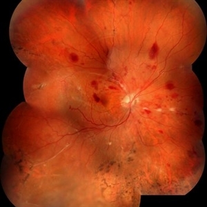
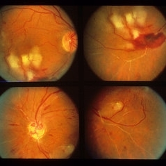
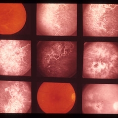
Color fundus photograph of the left eye of a 37-year-old man with Eales disease. Note the peripheral to mid-peripheral periphlebitis in multiple quadrants concurrently. Venous dilation and perivascular exudate can be observed. We can also note the demarcation between perfused and nonperfused retina.
Photographer: CBV- Eye Hospital Brasilia, DF, Brazil
Condition/keywords: Eales disease
-
 Eales Disease
Apr 3 2019 by Paola Brito, MD
8-year-old girl with positive Matoux test. She received laser in nasal retina. Peripheral vein occlusion, ischemic areas and neovascularization.
Photographer: Paola Brito, Hospital de la Luz, Mexico
Imaging device: retcam
Condition/keywords: Eales disease
-
 Eales Disease Causing TRD and Macular Edema in Pregnancy
Apr 21 2020 by Richard M Martindale, MD
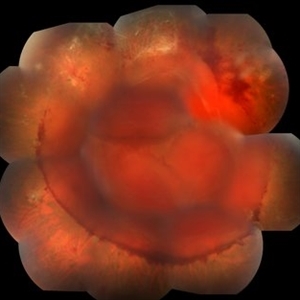
42-year-old pregnant African American with TRD and peripheral ischemia secondary to Eales disease. She was assigned this diagnosis of exclusion after a thorough work up for other identifiable causes of peripheral ischemia (e.g. sickle cell, syphilis, sarcoid, clotting disorders, SLE, TB, IP, FEVR). We elected to temporize her with PRP and Ozurdex in lieu of anti-VEGF medication given her pregnant status. Note: the Ozurdex pellet is visible in the inferior aspect of this photo.
Photographer: Retina Consultants of Alabama
Imaging device: Optos
Condition/keywords: Eales disease
-
 Sea Fan Neovascularisation
Apr 27 2015 by Neha Goel, MS DNB FRCS (Glasg)
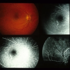
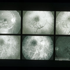
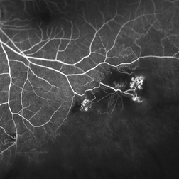
Fluorescein angiography of the left eye of a 40-year-old male.
Photographer: Neha Goel
Imaging device: Zeiss visucam
Condition/keywords: Eales disease, neovascularization elsewhere (NVE), vasculitis
-
 Disease of Eales
Aug 24 2017 by JEFFERSON R SOUSA, Tecg.º (Biomedical Systems Technology)
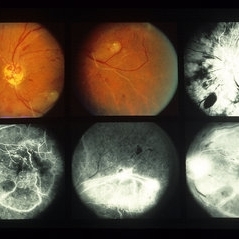
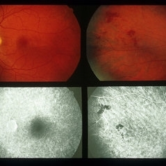
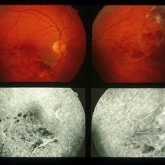
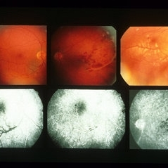
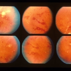
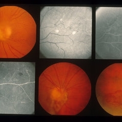
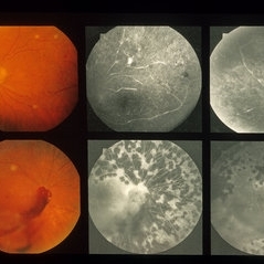
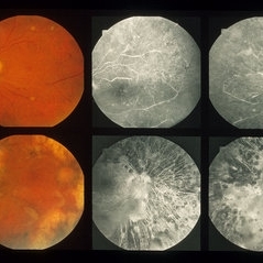
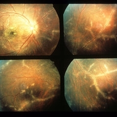
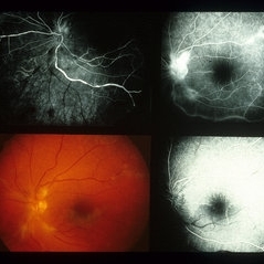
A 23-year-old male, Caucasian, attended the clinic with a complaint of progressive loss of vision. In the retinal mapping and retinography examination, we observed important alterations that suggested inflammatory processes. However, it turned negative for all laboratory tests.
Photographer: JEFFERSON R SOUSA - Study Center and Ophthalmological Research Dr. Andre M V Gomes, Institute Dr. Suel Abujamra São Paulo-Brazil
Imaging device: Topcon TRC-50 DX, Imaginet, campo de 50 graus. Flash 75 / Mosaic with 16 images.
Condition/keywords: Eales disease
-
 Disease of Eales
Aug 24 2017 by JEFFERSON R SOUSA, Tecg.º (Biomedical Systems Technology)
A 23-year-old male, Caucasian, attended the clinic with a complaint of progressive loss of vision. In the retinal mapping and retinography examination, we observed important alterations that suggested inflammatory processes. However, it turned negative for all laboratory tests.
Photographer: JEFFERSON R SOUSA - Study Center and Ophthalmological Research Dr. Andre M V Gomes, Institute Dr. Suel Abujamra São Paulo-Brazil
Imaging device: Topcon TRC-50 DX, Imaginet, campo de 50 graus. Flash 75 / Mosaic with 16 images.
Condition/keywords: Eales disease
-
 Eale's Diseas
-
 Eale's Disease
-
 Eale's Disease
-
 Eale's Disease
-
 Eale's Disease
-
 Eale's Disease
-
 Eale's Disease
-
 Eale's Disease
-
 Eale's Disease
-
 Eale's Disease
-
 Eale's Disease
-
 Eale's Disease
-
 Eale's Disease
-
 Eale's Disease
-
 Eale's Disease
-
 Eales Disease
Jan 31 2025 by Thirumalesh Mochi Basavaraj, MD
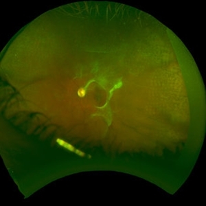
Ultra-wide field image of a 24 year old young healthy adult male with a visible sea fan neovascularization with partial PVD with vitreous and subhyaloid hemorrhage.
Photographer: Puttaswamy
Condition/keywords: Eales disease, sea fan, Ultra-wide field retinal imaging
-
 Eales Disease
Jan 31 2025 by Thirumalesh Mochi Basavaraj, MD
Ultra wide field image of a 24 year-old young healthy adult male with a visible sea fan neovascularization with partial PVD secondary to Scatter LASER photocoagulation with Vitreous and subhyaloid hemorrhage.
Photographer: Puttaswamy N K
Condition/keywords: Eales disease, Neovascularisation elsewhere (NVE), sea fan
-
 Eales Disease
Jul 11 2018 by Sarah Oelrich
Eales Disease
Photographer: Sarah Oelrich CRA, Southeastern Retina Associates, Knoxville TN
Imaging device: Optos 200tx
Condition/keywords: Eales disease
 Loading…