Search results (72 results)
-
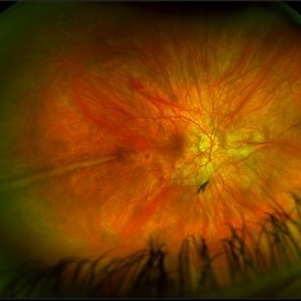
 IOL Drop
Dec 4 2025 by Surabhi Gupta, MS, DNB, FVRS
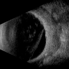
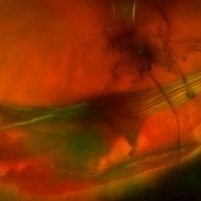
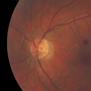
A 60 year old man presented with sudden dimunition of vision in right eye. His visual acuity was finger counting at 1 meter and best corrected visual acuity with +10 D was 6/9. Patient was diagnosed with spontaneous right eye IOL bag complex drop in vitreous cavity with superior HST and inferotemporal hole secondary to posterior vitreous detachment . Right eye montage color fundus photo shows rigid IOL bag complex in vitreous cavity with barraged superior HST and inferotemporal hole. Post barrage laser patient underwent pars plana vitrectomy with IOL explantation and scleral fixated IOL.
Photographer: Dr Surabhi Gupta
Imaging device: EDION FA
Condition/keywords: IOL drop
-
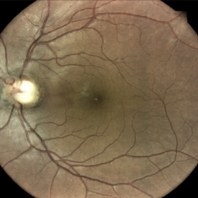
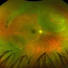
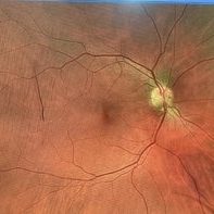 Posterior Vitreous Detachment
Sep 28 2025 by Sanauddin Samejo , Diploma (Ophthalmic Technician Training Course)
Posterior Vitreous Detachment (PVD)
Photographer: Sanauddin Samejo
Imaging device: Optos Silver Stone
Condition/keywords: posterior vitreous detachment, PVD
-
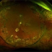
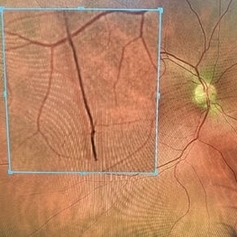 Posterior Vitreous Detachment
Sep 28 2025 by Sanauddin Samejo , Diploma (Ophthalmic Technician Training Course)
Posterior Vitreous Detachment (PVD)
Photographer: Sanauddin Samejo
Imaging device: Optos Silver Stone
Condition/keywords: posterior vitreous detachment, PVD
-
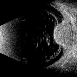 Choroidal Melanoma
Jul 3 2025 by Gustavo Uriel Fonseca Aguirre
This B-mode transverse ultrasound scan shows asteroid hyalosis with partial posterior vitreous detachment. A dome-shaped choroidal melanoma is observed in the inferior quadrant (preequatorial to equatorial region), appearing as a solid, regularly bordered lesion with heterogeneous internal structure and mild acoustic attenuation. Standardized A-mode reveals medium-to-low internal reflectivity. The tumor measures 11.62 mm in base diameter and 6.60 mm in height. The retina and choroid remain attached, with minimal suprachoroidal fluid in the inferior quadrant.
Photographer: Gustavo U. Fonseca Aguirre, Hospital Conde de Valenciana, Ciudad de México
Condition/keywords: choroidal melanoma
-
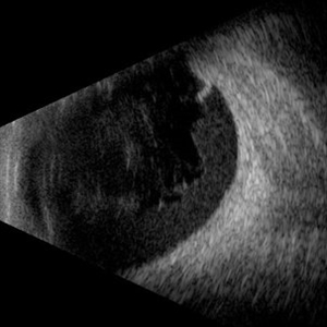 Hemorrhagic Vitreous Detachment
May 21 2025 by Gustavo Uriel Fonseca Aguirre
This B-mode longitudinal ultrasound scan shows a hemorrhagic vitreous detachment with the peripheral hyaloid strongly adherent to a retinal break. Associated vitreous and subhyaloid hemorrhage are present, indicating acute vitreoretinal traction.
Photographer: Gustavo U. Fonseca Aguirre, Hospital Conde de Valenciana, Ciudad de México
Condition/keywords: Hemorrhagic Vitreous Detachment
-
 Necrotizing Scleritis USG
Apr 17 2025 by Gustavo Uriel Fonseca Aguirre
This B-mode transverse ultrasound scan reveals necrotizing scleritis with inferior perilimbal uveal tissue prolapse, demonstrating scleral thinning and irregular uveal protrusion. Vitreous cellularity and partial vitreous detachment are also observed, indicating associated intraocular inflammation. These findings collectively characterize this severe inflammatory condition.
Photographer: Gustavo U. Fonseca Aguirre, Hospital Conde de Valenciana, Ciudad de México
Condition/keywords: necrotizing scleritis
-
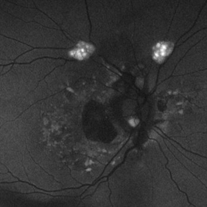 Astrocytic Hamartoma
Feb 27 2025 by Daniel Davis, OCT-C
Fundus autofluorescence photo of 55-year-old female with astrocytic hamartoma in association with tuberous sclerosis. No treatment options available, benign. Other findings include; Posterior Vitreous Detachment, Vitreous Hemorrhage, Hereditary Retinal Dystrophy, Vitreous Opacities, Hypertensive Retinopathy.
Photographer: Daniel Davis, OCT-C
Imaging device: Optos California
Condition/keywords: astrocytic hamartoma, fundus autofluorescence (FAF)
-
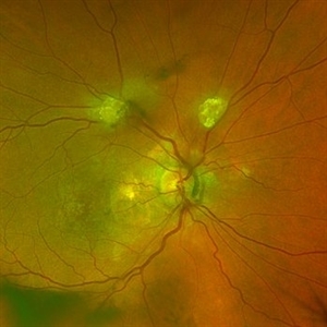 Astrocytic Hamartoma
Feb 27 2025 by Daniel Davis, OCT-C
Color fundus photo of 55-year-old female with Astrocytic Hamartoma in association with tuberous sclerosis. No treatment options available, benign. Other findings include; Posterior Vitreous Detachment, Vitreous Hemorrhage, Hereditary Retinal Dystrophy, Vitreous Opacities, Hypertensive Retinopathy.
Photographer: Daniel Davis, OCT-C
Imaging device: Optos California
Condition/keywords: color fundus photograph
-
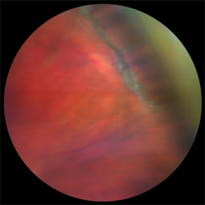
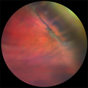
 Posterior Vitreous Detachment
Jan 31 2025 by Thirumalesh Mochi Basavaraj, MD
Intraoperative view of Triamcinolone-assisted posterior vitreous detachment.
Photographer: Thirumalesh Mochi Basavaraj
Condition/keywords: PVD induction, triamcinolone
-
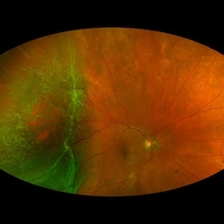 Rhegmatogenous Macula-On Retinal Detachment (Honeycomb)
Aug 6 2024 by Xitlali Caterina
Ultra-wide field fundus photograph of a 72 year old female with a macula-on retinal detachment with multiple breaks affecting her right eye. Patient presented in the office with flashes of light for five consecutive days prior. The patients vision was sc20/30 PHNI. The physician also noted an acute posterior vitreous detachment and lattice degeneration in the affect eye.
Photographer: Xitlali Caterina
Imaging device: Optos California RGB
Condition/keywords: honeycomb, lattice degeneration, Optos, posterior vitreous detachment, Retinal Detachment with Multiple Breaks, rhegmatogenous retinal detachment, ultra-wide field imaging
-
 Weiss Ring
Apr 22 2024 by SHIVANG CHAURASIA
Fundus photograph of a 68-year-old female with complaints of floaters.
Photographer: Dr SHIVANG CHAURASIA, GSVM MEDICAL COLLEGE, KANPUR, UTTAR PRADESH, INDIA
Imaging device: SMARTPHONE FUNDOSCOPY- IPHONE12
Condition/keywords: posterior vitreous detachment, Weiss ring
-
 Posterior Vitreous Detachment
Nov 1 2023 by ANKIT JAIN
USG B SCAN image showing membranous echoes with low to moderate spikes with free after movements with no attachment to disc suggestive of posterior vitreous detachment.
Photographer: DR ANKIT JAIN
Condition/keywords: B scan ultrasound, posterior vitreous detachment, PVD, ultrasound
-
 Vitreoretinal Traction with Adjacent Tear and Vitreous Hemorrhage
Oct 3 2023 by Alexis Singstock
Ultra-widefield fundus photograph of a 76 year old woman with vitreoretinal traction, an adjacent retinal tear and vitreous hemorrhage affecting the left eye. Patient was referred for retinal detachment and vitreous hemorrhage. Patient reports waking up the day prior to their appointment with "a lot of lines coming down the front, like swirling dirt in the left eye". Patient's vision was counting fingers at 1 ft. Dr. Joseph Boss noticed a horseshoe tear inferior to traction on exam and with the help of ultra-widefield imaging. Dr. Boss performed laser retinopexy to tear and impending tear at site of traction. Patient is scheduled for pars plana vitrectomy for dense vitreous hemorrhage.
Photographer: Alexis Singstock
Imaging device: Optos California
Condition/keywords: acute posterior vitreous detachment, fundus photography, left eye, Optos, OPTOS CALIFORNIA, pseudocolor, ULTRA WIDE FIELD, vitreoretinal traction, vitreous hemorrhage
-
 Evolving Weiss Ring
Sep 11 2022 by Michael B Green, MD, MBA
Fundus photograph of a 62-year-old female with an evolving Weiss-ring in the process of separating from the optic disc.
Condition/keywords: posterior vitreous detachment, PVD, Weiss ring
-
 Vitreous Base Avulsion
Feb 21 2022 by Maxwell J Wingelaar, MD
24-year-old male with a vitreous base avulsion with a history of blunt force trauma to the eye
Condition/keywords: vitreous detachment
-
 Vitreous Base Avulsion
Feb 21 2022 by Maxwell J Wingelaar, MD
24-year-old male with a vitreous base avulsion with a history of blunt force trauma to the eye
Condition/keywords: vitreous detachment
-
 Prominent Long Ciliary Nerve
Jan 25 2022 by Kachelle Brown
Ultra-wide field photograph of a 48-year-old female with a prominent long ciliary nerve. Patient presented asymptomatic, and was referred for a macula on retinal detachment. Patient was diagnosed with high myopia and a posterior vitreous detachment, and the physician discussed increased risk of floaters, myopic degeneration and retinal detachment associated with high myopia. -24.00 prior to cataract surgery OU per patient.
Photographer: Kachelle Brown
Imaging device: Optos California
Condition/keywords: fundus photograph, high myopia, long ciliary nerve, optos, right eye, ultra-widefield image
-
Macular Hematoma Secondary Valsalva Maneuver
Oct 14 2021 by Islam bechakh
A 32-year-old man, who has presented for 02 months, a macular hematoma secondary to a Valsalva maneuver. He benefited from an attempt to open the hematoma with a Yag laser, but to no avail. We operated on and performed a 23G vitrectomy with posterior vitreous detachment, and discovered an epiretinal membrane which separated the hematoma from the posterior hyaloid. After removal of this membrane and aspiration of red blood cells and fibrin, the macula regained a normal appearance with good functional recovery.
Photographer: Islam Bechakh
Condition/keywords: epiretinal membrane (ERM), ERM, Macular hematoma, Valsalva maneuver
-
 Sea-Saw Vitreoretinal Dance
Mar 12 2021 by RUSHIK PATEL
Fundus photograph of 50-year-old female showing posterior vitreous detachment, 2 retinal tears with localized retinal detachment exactly 180 degrees apart with optic disc in between as a Falcrum giving an appearance of sea saw retinal tears. Macula was attached with lattice retinal degeneration superiorly.
Photographer: Rushik Patel, Netralaya Super Speciality Eye Hospital
Condition/keywords: peripheral lattice degeneration, retinal tear
-
 Scleral Buckle
Dec 31 2020 by Tammy Mclaughlin
Central serous chorioretinopathy OS (Incidental SRF 6/25/20 a little worse then stable). Repaired recurrent retinal detachment with PVR OS (new inferior starfold, anterior loop, and heavy pigment. s/p PPV 11/19/19, PPV/SBP/MP/SO 1/7/20). Posterior vitreous detachment OD. Floaters OD. Epiretinal membrane OS (mild).
Photographer: Tammy Mclaughlin, Carolina Retina Center, Sumter SC
Condition/keywords: scleral buckle
-
 Posterior Vitreous Detachment
Sep 1 2020 by J. Sebag, MD, FACS, FRCOphth, FARVO
Left: Preset lens biomicroscopy of PVD in the left eye of a subject with a widely dilated pupil. The detached posterior vitreous cortex is seen (arrows) as is the optic disc and retinal vasculature (upper left). (courtesy of C. L. Trempe MD, Harvard Medical School, Boston, MA) [Sebag J: Vitreous – in Health & Disease Springer, New York, 2014; image © Springer Nature, reprinted with permission] Right: B-scan ultrasonography of PVD images the detached posterior vitreous cortex with a visible Weiss Ring.
Condition/keywords: posterior vitreous detachment
-
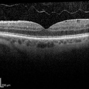 OCT of a Posterior Vitreous Detachment
Nov 26 2019 by Geoffrey G. Emerson, MD, PhD, FASRS
OCT of a posterior vitreous detachment.
Condition/keywords: optical coherence tomography (OCT), posterior vitreous detachment
-
 Weiss Ring
Oct 22 2019 by Jessica Norkus
Fundus photo taken on TopCon TRC 50Dx camera of a 60-year-old patient who has experienced an acute PVD. Chief complaint of "large floater" OS prompted exam. Physician noted a significant Weiss Ring and requested fundus color photo for documentation.
Photographer: Jessica Norkus, COA (Retina Specialists of Michigan)
Imaging device: TopCon TRC 50Dx
Condition/keywords: color fundus photograph, color photo, fundus photograph, nerve, optic disc, posterior vitreous detachment, retina, vitreous floaters, Weiss ring
-
 Weiss Ring
Apr 8 2019 by Gary R. Cook, MD, FACS
Weiss ring ahead of optic disc following a recent posterior vitreous detachment (PVD)
Imaging device: Topcon VT-50
Condition/keywords: posterior vitreous detachment, Weiss ring
-
 Posterior Vitreous Detachment
Mar 21 2019 by Michael Politis, MD
Intra-op image of a PVD induction using Kenalog in a retinal detachment after retained nuclear material
Photographer: Michael Politis MD, McGill University, Montreal, Canada
Imaging device: Karl Stroze
Condition/keywords: kenalog, PVD induction
 Loading…