Search results (206 results)
-
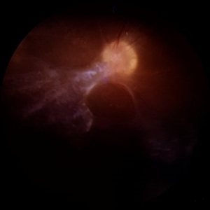
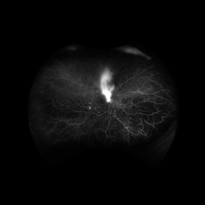
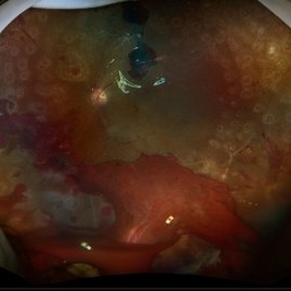
 Napkin on my Retina: The Diabetic TRD
Nov 17 2025 by SHRADDHA RAJ SHRIVASTAVA
Left eye pseudocolor widefield fundus image of a 57 year old patient with poorly controlled diabetes mellitus, presenting with BE tractional retinal detachment (TRD) on a background of high risk proliferative diabetic retinopathy (PDR). The disc, macula and posterior pole anatomy is obscured and distorted by the extensive tractional fibrovascular component, giving the appearance of a crumpled napkin on this patient's retina.
Photographer: Dr. Shraddha Raj Shrivastava
Imaging device: Nidek Mirante SLO/OCT (Confocal scanning/Spectral domain OCT)
Condition/keywords: Diabetic Tractional Detachment, Diabetic Tractional Retinal Detachment involving the Macula, PDR, proliferative diabetic retinopathy (PDR), tractional retinal detachment
-
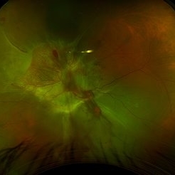
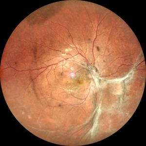
 Napkin on my Retina: The Diabetic TRD
Nov 17 2025 by SHRADDHA RAJ SHRIVASTAVA
Left eye pseudocolor fundus image of a 57 year old patient with poorly controlled diabetes mellitus, presenting with BE tractional retinal detachment (TRD) on a background of high risk proliferative diabetic retinopathy (PDR). The disc, macula and posterior pole anatomy is obscured and distorted by the extensive tractional fibrovascular component, giving the appearance of a crumpled napkin on this patient's retina.
Photographer: Dr. Shraddha Raj Shrivastava
Imaging device: Nidek Mirante SLO/OCT (Confocal scanning/Spectral domain OCT)
Condition/keywords: Diabetic Tractional Detachment, Diabetic Tractional Retinal Detachment involving the Macula, proliferative diabetic retinopathy (PDR), tractional retinal detachment
-
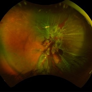
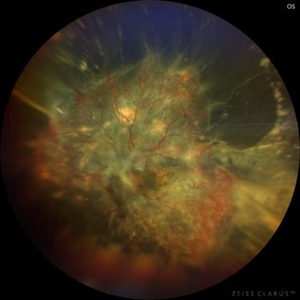
 Fibrovascular Fortress: Disc in Captivity
Nov 4 2025 by SHRADDHA RAJ SHRIVASTAVA
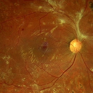
Right eye pseudocolor fundus photo of 67 year old male, diagnosed with both eyes proliferative diabetic retinopathy, showing extensive neovascularisation at the disc (NVD), with table-top configuration of the diabetic tractional retinal detachment (TRD) obscuring the view of the disc and threatening the macula. We can also see a mound of pre-retinal bleed and active neovascular frond nasal to the disc.
Photographer: Dr. Shraddha Raj Shrivastava
Imaging device: Nidek Mirante SLO/OCT (Confocal scanning/Spectral domain OCT
Condition/keywords: Active PDR Tractional retinal Detachment, Diabetic Tractional Detachment, Neovascularisation at the Disc (NVD), proliferative diabetic retinopathy (PDR), TABLE TOP TRD, tractional retinal detachment
-
 Vasoproliferative Tumor
Oct 27 2025 by Virginia Gebhart
34 year old male with retinal vasoproliferative tumor with traction detachment temporally and striae in the macula. Recommend vitrectomy prior to cryotherapy to release vitreous from retina. BCVA 20/30
Photographer: Virginia Gebhart, Retina Consultants of Carolina
Imaging device: Optos California
Condition/keywords: traction retinoschisis, tractional retinal detachment, Vasoproliferative Tumor
-
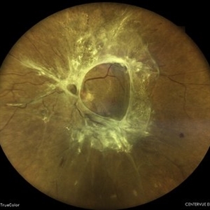
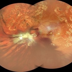 Table Top Tractional Retinal Detachment With Vitreous Hemorrhage in a Case of Proliferative Diabetic Retinopathy
Sep 12 2025 by Akansha Sharma
Color fundus photograph of a 56 year old male with table top tractional retinal detachment with vitreous hemorrhage in a case of proliferative diabetic retinopathy.
Photographer: DR. AKANSHA SHARMA
Condition/keywords: pan-retinal photocoagulation (PRP), PDR, proliferative diabetic retinopathy (PDR), PRP, TABLE TOP TRD, tractional retinal detachment, TRD, VH, vitreous hemorrhage
-
 Macular Hole Due to Proliferative Diabetic Retinopathy
Aug 13 2025 by Ricardo Leitão Guerra
A macular hole formation after anti-VEGF injection prior to vitrectomy for tractional retinal detachment in a patient presenting proliferative diabetic retinopathy.
Photographer: Ricardo Leitão Guerra
Imaging device: ZEISS CLARUS 700
Condition/keywords: macular hole, proliferative diabetic retinopathy (PDR)
-
 Unstable PDR s/p Laser
Aug 4 2025 by Anjana Mirajkar, MS Ophthalmology
Fundus photograph of a 60 year old male with an unstable PDR showing traction at the posterior pole with sub hyaloid hemorrhage. Peripheral PRP marks can be seen.
Photographer: Dr. Anjana Mirajkar- HV Desai eye hospital ,Pune
Imaging device: Optos
Condition/keywords: pan-retinal photocoagulation (PRP), proliferative diabetic retinopathy (PDR), subhyaloid hemorrhage, tractional retinal detachment
-
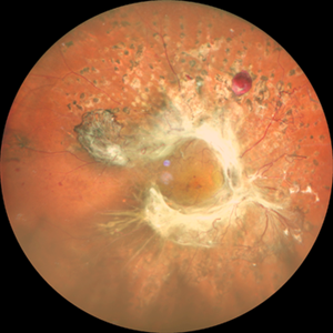
 Annular Tractional Retinal Detachment
Jul 5 2025 by César Adrián Gómez Valdivia, MD
Fundus photograph of an 66 YO female patient diagnosed with advanced proliferative diabetic retinopathy.
Photographer: @eyemissu2
Imaging device: TOPCON TRC-50DX
Condition/keywords: tractional retinal detachment
-
 Tractional Retinal Detachment
Jun 4 2025 by Paulina Araujo
The 55-degree central fundus photograph of the right eye reveals a thickened and opacified hyaloid exerting traction on the optic disc and posterior pole of the retina, along with hard exudates and microaneurysms consistent with advanced proliferative diabetic retinopathy.
Photographer: Paulina D.Araujo Martínez, Asociación para Evitar la Ceguera en México I.A.P., Hospital Dr Luis Sánchez Bulnes.
Condition/keywords: tractional retinal detachment
-
 Tractional/Rhegmatogenous Retinal Detachment
May 29 2025 by Jenn Geelan
48 year-old male with a combined Tractional/ Rhegmatogenous retinal detachment with NVD and Persistent Fetal Vasculature in the left eye.
Photographer: Jenn Geelan, Retina-Vitreous Surgeons of CNY
Condition/keywords: fundus photograph, Neovascularisation at the Disc (NVD), OPTOS CALIFORNIA, persistent fetal vasculature (PFV), retinal detachment, rhegmatogenous retinal detachment, tractional retinal detachment
-
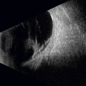
 Advanced Proliferative Diabetic Retinopathy
Apr 9 2025 by Gustavo Uriel Fonseca Aguirre
B-mode ultrasound of a patient with long-standing poorly controlled diabetes demonstrates characteristic findings of advanced proliferative diabetic retinopathy. The examination reveals moderate vitreous hemorrhage appearing as diffuse hyperechoic opacities throughout the vitreous cavity, along with a posterior hyaloid membrane densely infiltrated by hemorrhagic material, showing irregular thickening and increased reflectivity. A mild subhyaloid hemorrhage is visible as a subtle hyphema-like space anterior to the retinal surface. The study documents a total tractional retinal detachment, evidenced by rigid retinal folds with clear insertion points of vitreous strands, accompanied by a significant subretinal hemorrhage seen as a prominent hyperechoic collection beneath the elevated retina. These findings collectively illustrate the severe vitreoretinal interface pathology characteristic of end-stage diabetic eye disease, with predominant tractional components and distinct echographic stratification of hemorrhagic layers - from anterior vitreous involvement to deeper subretinal blood accumulation.
Photographer: Gustavo U. Fonseca Aguirre, Hospital Conde de Valenciana, Ciudad de México
Condition/keywords: diabetic retinopathy, tractional retinal detachment, Vitreous hemorrhage
-
 Macular Hole in Tractional Retinal Detachment
Apr 1 2025 by Gustavo Uriel Fonseca Aguirre
B-scan findings in a diabetic patient reveal vitreous hemorrhage, blood-soaked hyaloids, and tractional retinal detachment with an associated macular hole in the posterior pole.
Photographer: Gustavo U. Fonseca Aguirre, Hospital Conde de Valenciana, Ciudad de México
Condition/keywords: macular hole, tractional retinal detachment
-
 Combined Traction and Rhegmatogenous Retinal Detachment From Proliferative Diabetic Retinopathy
Mar 27 2025 by Nikhil K Bommakanti, MD
A middle-aged patient presented with a combined traction and rhegmatogenous retinal detachment.
Condition/keywords: Active PDR Tractional retinal Detachment, PDR, Retinal Detachment, rrd, TRD
-
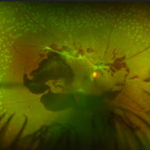
 Diabetic Tractional Retinal Detachment Involving the Macula OD
Feb 21 2025 by Kaitlyn Anderson
57-year-old female. Diabetic Tractional Retinal Detachment involving the Macula OD. Active Proliferative Diabetic Retinopathy
Photographer: Kaitlyn Anderson TN Retina Nashville TN
Imaging device: Optos Fluorescein Angiogram
Condition/keywords: Active PDR Tractional retinal Detachment
-
 Diabetic Tractional Retinal Detachment
Jan 6 2025 by Kavitha Duraipandi, MD DNB FICO FRCS
55 year old patient , with poor metabolic control , came with right eye gradual loss of vision. Patient had partial inadequate retinal laser in the past. Funds examination showed fibrovascular proliferation over the arcades with tractional retinal detachment. Patient under went right eye Pars plans vitrectomy with endo laser with silicone oil injection. patient was given pre op anti VEGF injection.
Condition/keywords: TRD
-
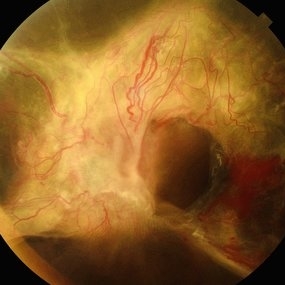
 Extramacular TRD in Idiopathic Occlusive Vasculitis
Dec 5 2024 by Tejaswita Verma
Fundus photo showing extramacular TRD in a 16 year old boy with idiopathic occlusive vasculitis secondary to presumed IOTB. History of taking ATT for 6 months , Mantoux positive previously. Vision was 6/6P,other eye had funnel RD .
Photographer: DR. TEJASWITA VERMA
Imaging device: MIRANTE
Condition/keywords: tractional retinal detachment, vasculitis
-
 Combined Pathology
Oct 26 2024 by rahul saradge
53 year old male patient was presented with a complaints of diminished vision in LE since 1 month. The BCVA in RE was 6/36p and LE was CF 1/2m. Ocular dilated examination showed RE temporal CD with ?CRVO,OIS and OS showed TRD and old Hemi CRVO. Patient was injected with PST tricot followed by PRP laser at an interval of 1 week. Patient improved to BCVA 6/9.
Photographer: Aishwarya Bangar Isha Netralaya Thane
Imaging device: optos
Condition/keywords: choroidal detachment, crvo, ois, optos, pan retinal photocoagulation, tractional retinal detachment
-
 Combined Retinal Detachment With Macular Hole
Sep 28 2024 by Tejaswita Verma
Fundus image of the LE of a 67 year old diabetic, hypertensive female with CF 3metres vision showing combined RD with FTMH, in a pseudophakic eye. She was lost to follow up status post 2 anti VEGF injections received 8 months back due to typhoid fever.
Photographer: DR. TEJASWITA VERMA
Imaging device: MIRANTE
Condition/keywords: full thickness macular hole, proliferative diabetic retinopathy (PDR), tractional retinal detachment
-
 Tractional Detachment of Retina
Aug 21 2024 by Jordyn Beckman
18 year old male with tractional detachment of Retina, chronic macular hole and silicone oil s/p RD repair x2. BCVA CF @2 ft, fellow eye prosthetic.
Photographer: Jordyn Beckman
Imaging device: Optos California
Condition/keywords: Macular hole, preretinal fibrosis, Retinal Detachment, scleral buckle, silicone oil, TRACTION, tractional retinal detachment
-
 Diabetic Tractional Retinal Detachment 1 week s/p SO fill
Aug 14 2024 by Virginia Gebhart
21 year old male 1 week s/p PPV/laser/STR/SO. Eye is stable, PRHs inferior and superior, possible traction from PRH/membrane. Will observe and let clot liquify, will consider scleral buckle if no improvement
Photographer: Virginia Gebhart
Imaging device: Optos California
Condition/keywords: Diabetic Tractional Detachment, retinal detachment of the macula, silicone oil
-
 New Vessels
Aug 12 2024 by BENJAMIN ABOYTES RIOS
Fundus photograph of an 34-year-old woman with a tractional retinal detachment secondary to diabetic retinopathy.
Photographer: Benjamín Aboytes Ríos, Asociación Para Evitar la Ceguera en México, Ciudad de México.
Imaging device: Clarus 700
Condition/keywords: Diabetes, diabetic retinopathy, Diabetic Tractional Retinal Detachment involving the Macula
-
 Tractional Retinal Detachment in a Case of Proliferative Diabetic Retinopathy
Aug 6 2024 by Akansha Sharma
Color fundus photograph of a 60 year old female with tractional retinal detachment in a case of proliferative diabetic retinopathy.
Photographer: Dr. Akansha Sharma, Bharati Eye Hospital
Condition/keywords: fibrovascular proliferation, PDR, Proliferative Diabetic retinopathy, tractional retinal detachment, TRD
-
 Annular Tractional Retinal Detachment
Jul 4 2024 by Hector Gabriel Moreno Solano, MD, MHA
52-year-old Hispanic female patient with a diagnosis of type II diabetes mellitus of 15 years of evolution, comes to the retina service for progressive visual loss in the right eye (single functional eye) with visual acuity of 20/100, Fundus examination reveals laser-modified proliferative diabetic retinopathy with activity + annular tractional retinal detachment with macular involvement.
Photographer: Hector Gabriel Moreno Solano, MD, MHA, HGZ #20 IMSS Puebla.
Imaging device: Mirante
Condition/keywords: macular detachment, proliferative diabetic retinopathy (PDR), tractional retinal detachment
-
 Post Combined Surgery of Cataract, TRD & Vitreous Hemorrhage
Jun 27 2024 by Sanauddin Samejo , Diploma (Ophthalmic Technician Training Course)
A 27 year-old diabetic female visited the clinic one week after combined surgery of cataract, tractional retinal detachment and vitreous hemorrhage.
Photographer: Sanauddin Samejo, Burjeel Hospital, Abu Dhabi, UAE
Imaging device: Silver Stone Optos
Condition/keywords: Combined Surgery Cataract Tractional Retinal Detachment Vitreous Hemorrhage, POST SURGERY, Retinal Detachment, TRD
-
 Cataract, Tractional Retinal Detachment, Vitreous Hemorrhage
Jun 27 2024 by Sanauddin Samejo , Diploma (Ophthalmic Technician Training Course)
A 27 years Female visited Dr Madhav Rao's Clinic with Bilateral Cataract, Tractional Retinal Detachment and Vitreous Hemorrhage, she is Diabetes.
Photographer: Sanauddin Samejo, Burjeel Hospital, Abu Dhabi, UAE
Imaging device: Optos Silver Stone
Condition/keywords: cataract, Retinal Detachment with Vitreous Hemorrhage, TRD
 Loading…