Search results (74 results)
-
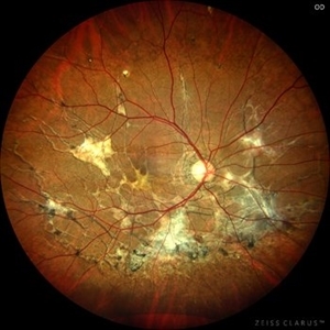
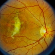
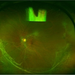
 Serpiginous Choroidopathy
Jun 23 2025 by César Adrián Gómez Valdivia, MD
Fundus photograph of a 29 year-old female patient diagnosed with Serpiginous Choroidopathy. Finings were bilateral. The most common complication of SC is choroidal neovascularization affecting up to 35% of patients. Other reported complications are subretinal fibrosis, cystoid macular edema, branch vein occlusion, serous retinal detachment, optic disc neovascularization ,and anterior uveitis.
Photographer: @eyemissu2
Imaging device: TOPCON TRC-50DX
Condition/keywords: serpiginous choroiditis
-
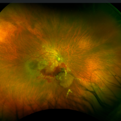
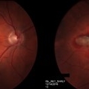
 Serpiginous Choroidopathy
Jun 23 2025 by César Adrián Gómez Valdivia, MD
Fundus photograph of a 29 year-old female patient diagnosed with Serpiginous Choroidopathy. Finings were bilateral. The most common complication of SC is choroidal neovascularization affecting up to 35% of patients. Other reported complications are subretinal fibrosis, cystoid macular edema, branch vein occlusion, serous retinal detachment, optic disc neovascularization, and anterior uveitis.
Photographer: @eyemissu2
Imaging device: California ICG OPTOS
Condition/keywords: serpiginous choroiditis
-
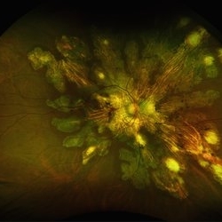
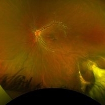
 VKH Pseudotumor – Chronic Subretinal Fibrosis
May 11 2025 by Felipe Murati
Ultra-widefield fundus image from a 36-year-old woman with chronic VKH syndrome showing a pseudotumor-like subretinal fibrotic lesion in the right eye. The lesion developed after multiple relapses and remained stable over a 1-year follow-up with immunosuppressive treatment including prednisone, mycophenolate mofetil, and adalimumab. No active choroiditis or exudative detachment was observed. Multimodal imaging was essential for disease monitoring.
Photographer: Felipe A. Murati, MD, University of Arizona
Imaging device: Optos California ultra-widefield retinal imaging system, single-capture, color fundus modality.
Condition/keywords: adalimumab, chronic inflammation, granulomatous uveitis, OCT, Optos ultra-widefield imaging, pseudotumor, subretinal fibrosis, VKH, Vogt-Koyanagi-Harada
-
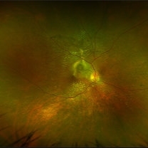
 VKH Pseudotumor – Fluorescein Angiography
May 11 2025 by Felipe Murati
Fluorescein angiography image from a 36-year-old woman with chronic Vogt-Koyanagi-Harada (VKH) syndrome showing a pseudotumor-like lesion with late-phase staining and no active leakage. The image highlights subretinal fibrosis in the right eye, stable under long-term immunosuppressive therapy with mycophenolate mofetil and adalimumab. No signs of active choroiditis are present, confirming a quiescent phase.
Photographer: Felipe A. Murati, MD, University of Arizona
Imaging device: Optos California, fluorescein angiography modality
Condition/keywords: choroiditis, Fluorescein angiography, granulomatous uveitis, Optos FA, pseudotumor, subretinal fibrosis, VKH, Vogt-Koyanagi-Harada
-
 Subretinal Fibrosis
Jan 14 2025 by Kimberly Wakester
Fundus photograph of an 86-year-old woman with the end stage of Age-related Macular Degeneration in the left eye. Patient went unseen for 3-4 years prior to establishing care at our practice. Due to the significant amount of subretinal fibrosis, treatment was not recommended due to limited visual recovery. Patient was advised of monocular vision and the importance of follow up care.
Photographer: Kimberly Wakester, COA
Imaging device: Optos California
Condition/keywords: AMD, subretinal fibrosis
-
 Pseudoxanthoma Elasticum Associated Angioid Streaks
Aug 18 2024 by KANWALJEET HARJOT MADAN, M.S. (Ophthalmology); FAICO (Vitreous - Retina)
This is fundus photograph of a young 31 years male patient depicting Angioid streaks emanating from optic nerve towards the periphery and subretinal fibrosis. There is peau de orange appearance temporal to fovea with Salmon Spots in periphery. He was diagnosed to have Pseudoxanthoma Elasticum.
Photographer: Dr. Kanwaljeet Harjot Madan, M.S. (Ophthalmologist) Fellow in Vitrous & Retina. Thind Eye Hospital, Jalandhar City. Punjab. India
Imaging device: Zeiss Clarus
Condition/keywords: Angioid Streaks, fundus photograph, pseudoxanthoma elasticum (PXE)
-
 Ocular Toxocariasis
Jul 4 2024 by Brandon I Fram, MD, BS
4 yo with toxocariasis-related peripheral granuloma with adhesion to the macula and macular subretinal fibrosis. Positive Toxocara titers.
Condition/keywords: toxocara canis, toxocara granuloma, toxocariasis
-
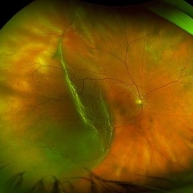 Retinal Detachment
Mar 28 2024 by Virginia Gebhart
68 year male with chronic appearing retinal detachment with subretinal bands and subretinal fibrosis. Demarcation line present, SRF splits the fovea on OCT.
Photographer: Virginia Gebhart
Imaging device: Optos California
Condition/keywords: chronic retinal detachment, Retinal Detachment
-
 WED-ARMD-CNVM
Sep 29 2023 by PUSHPANJALI BADOLE
Fundus photograph of a 72 year old male with right eye choroidal neovascularization. There is a thick membrane over macula along with subretinal fibrosis in right eye. Left eye shows drusen.
Photographer: NITIN DESALE, ISHA NETRALAYA, KALYAN
Imaging device: DAYTONA OPTOS
Condition/keywords: choroidal neovascular membrane (CNVM), choroidal neovascularization (CNV)
-
 Subretinal fibrosis
Sep 14 2023 by Ben Serar
Fundus photograph of LE showing a scarred lesion at the macula, with sub retinal fibrosis.
Condition/keywords: macular scar, Subretinal fibrosis
-
 Subretinal fibrosis
Sep 12 2023 by Ben Serar
Fundus photograph of RE showing scarring at the macula with subretinal fibrosis
Condition/keywords: Subretinal fibrosis
-
 Sunset Glow Fundus
May 15 2022 by Manuel Ángel Alcántara Delgado, MD
Optomap ultra-widefield retinal imaging of an 35-year-old woman showed sunset glow fundus, multiple nummular chorioretinal atrophic lesions, macular subretinal fibrosis and pigment clumping in chronic recurrent stage of Vogt-Koyanagi-Harada disease.
Photographer: Manuel Ángel Alcántara Delgado. Conde de Valenciana.
Condition/keywords: abnormal retina, benign pigmented lesions, pigment clumps, retinal fibrosis, uveitis, Vogt-Koyanagi-Harada
-
 Neovascular Age-Related Macular Degeneration (1)
Apr 28 2021 by Ambar Faridi, MD
80-year-old woman with neovascular age-related macular degeneration with large subretinal hemorrhage, hemorrhagic PED, and vascular lipid exudation.
Photographer: Jennifer Tu-Bui, VA Portland Health Care System
Condition/keywords: subretinal fibrosis, subretinal hemorrhage
-
 Blunt Ocular Trauma Due to Firework Injury
Jun 9 2020 by Brittany Rota
Ultra- widefield pseudocolor image of an 18-year-old male with blunt ocular trauma in the right eye due to a firework injury. The patient presented with commotio retinae (sclopteria), an acute vitreous hemorrhage, choroidal rupture, and a subretinal hemorrhage. The referring physician performed surgery on the lateral rectus muscle which was macerated but not severed, and several orbital fibrous foreign bodies were removed from the posterior orbit. The globe was intact. There is no evidence of retinal tear in the region of sclopetaria; however, there is complete necrosis of the temporal peripheral choroid and retina. The vitreous hemorrhage was slowly clearing on his exam 6-9-2020. The patient is developing subretinal fibrosis. The physician is concerned about the choroidal rupture that is visible through the submacular hemorrhage. There is one rupture that appears to course directly under the fovea. The physician states that if this is the case, his vision most likely will be 20/200 or worse. His vision was hand motion in all fields except nasally, which he was unable to see hand motion at his visit on 6-9-2020.
Photographer: Brittany Rota
Imaging device: Optos California
Condition/keywords: blunt trauma, choroidal rupture, commotio retinae, fibrosis, firework injury, fundus photograph, hand motion, necrotizing retina, Optos, pseudocolor, subretinal hemorrhage, vitreous hemorrhage
-
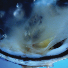 Subretinal Fibrosis and Uveitis Syndrome
May 18 2020 by McGill University Health Centre
Uveitis syndrome is a rare posterior uveitis that usually begins as a multifocal choroiditis and then progresses to subretinal fibrosis. Recurrences are not uncommon and the visual prognosis is generally poor. In this enucleation specimen, a thickened choroid is clearly observed (arrow). The retina is detached and a fibrovascular subretinal membrane is present (arrowhead).
Condition/keywords: subretinal fibrosis, uveitis
-
 Subretinal Fibrosis (PPCNVM and POHS) OS
Sep 18 2019 by John S. King, MD
57-year-old white male with history of PPCNVM OS and POHS OU here for a routine visit. History of avastin in 2014, and stable since then. Va OS 20/20. PP scar with macular subretinal fibrosis. No heme or exudates. CR spot supero-nasally.
Photographer: Shelly Blair
Imaging device: Topcon 50
Condition/keywords: choroidal neovascular membrane (CNVM), ocular histoplasmosis syndrome (OHS), peripapillary choroidal neovascularization (PPCNVM), presumed ocular histoplasmosis syndrome (POHS)
-
 Chorioretinal Scars with Subretinal Fibrosis and an old Retinal Detachment
May 3 2018 by Nichole Lewis
Chorioretinal scars with subretinal fibrosis and an old retinal detachment.
Photographer: Nichole Lewis
Condition/keywords: chorioretinal scar, chronic retinal detachment, subretinal fibrosis
-
 Wagner Syndrome
Aug 1 2017 by Eitae Kim, MD
Ulltra wide field fundus photograph of 19-year-old male with Wagner syndrome which shows peripheral subretinal fibrosis and pigmentary degeneration.
Photographer: Eitae Kim, BOIM retinal center, Pureun eye hospital
Condition/keywords: subretinal fibrosis, ultra-wide field imaging, Wagner disease
-
 Subretinal Thickening and Subretinal Hemorrhage – Stereo Color Fundus Photograph
Mar 9 2017 by James B. Soque, CRA, OCT-C, COA, FOPS
Color fundus stereo photograph of a 52-year-old white male with VA loss to 20/200 of unknown etiology. Dilated fundus examination of the right eye reveals a fibrotic scar with subretinal thickening and subretinal hemorrhage.
Photographer: James B Soque, CRA, OCT-C, COA
Imaging device: Topcon TRC 50 DX, MERGE Software
Condition/keywords: blood, color fundus photograph, color photo, stereo pair, subretinal blood, subretinal fibrosis, subretinal thickening
-
 Peculiar Acute Subretinal Fibrosis
Feb 24 2015 by David Callanan, MD
Peculiar acute subretinal fibrosis.
Condition/keywords: subretinal fibrosis
-
 Peculiar Acute Subretinal Fibrosis
Feb 24 2015 by David Callanan, MD
Peculiar acute subretinal fibrosis.
Condition/keywords: subretinal fibrosis
-
 Peculiar Acute Subretinal Fibrosis
Feb 24 2015 by David Callanan, MD
Peculiar acute subretinal fibrosis.
Condition/keywords: subretinal fibrosis
-
 Peculiar Acute Subretinal Fibrosis
Feb 24 2015 by David Callanan, MD
Peculiar acute subretinal fibrosis.
Condition/keywords: subretinal fibrosis
-
 Peculiar Acute Subretinal Fibrosis
Feb 24 2015 by David Callanan, MD
Peculiar acute subretinal fibrosis.
Condition/keywords: subretinal fibrosis
-
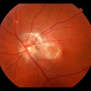
 Subretinal Fibrosis
Feb 20 2015 by H. Michael Lambert, MD
Posterior pole with possible subretinal fibrosis.
Condition/keywords: subretinal fibrosis
 Loading…