Search results (206 results)
-
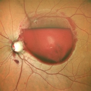
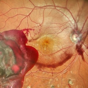
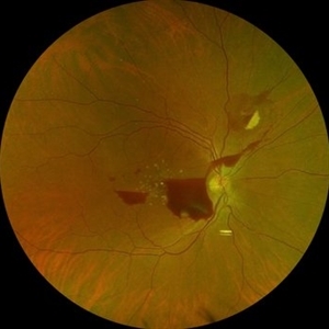
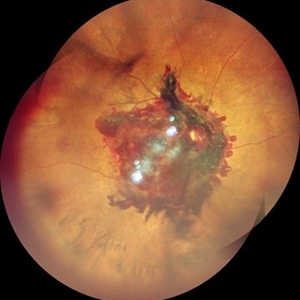 Subhyaloid Hemorrhage With Vitreous Hemorrhage
Sep 12 2025 by Akansha Sharma
Color fundus photograph of a 56 year old hypertensive and diabetic female who presented with subhyaloid hemorrhage along with vitreous hemorrhage after being administered high dose anti-platelet therapy pre- and post a cardiac procedure.
Photographer: DR. AKANSHA SHARMA
Condition/keywords: SHH, sub ILM hemorrhage, subhyaloid hemorrhage, VH, vitreous hemorrhage
-
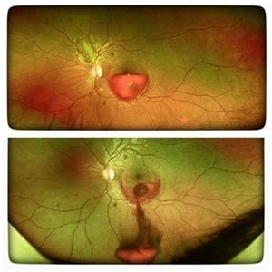 YAG Laser Hyaloidotomy
Aug 31 2025 by Giriraj Vibhute
A 24-year-old young man presented with sudden loss of vision in left eye following history of rigorous coughing. Visual acuity in RE was 6/6, LE was 6/60p. Fundoscopy showed bilateral multiple small intraretinal hemorrhages with LE large premacular subhyaloid hemorrhage just covering the fovea suggestive of bilateral valsalva retinopathy changes. Nd:YAG laser hyaloidotomy was performed to left eye the same day (A250; 2mJ;6 SHOTS). Visual acuity improved to 6/9 immediately following the procedure. After 1 week, the subhyaloid hemorrhage had completely cleared with dispersed intragel hemorrhage in the inferior vitreous cavity with visual acuity of 6/6 in left eye
Photographer: Dr Vani S. MM Joshi eye institute, Hubli
Condition/keywords: valsalva retinopathy, YAG HYALOIDOTOMY
-
 Premacular Hemorrhage Secondary to Recreational Laser Exposure
Aug 22 2025 by Carlos Valdez Prado
A 40-year-old male with accidental exposure to a recreational laser during a party presents with sudden visual loss following the laser exposure. On examination, a premacular hemorrhage with a double-ring sign is observed in the macular region.
Photographer: Dr. Carlos A. Valdez Prado, Hospital Militar de Especialidades Oftalmologicas
Imaging device: Optos
Condition/keywords: Hemorrhage, premacular hemorrhage, Retina, subhyaloid hemorrhage, sublimitante
-
 Unstable PDR s/p Laser
Aug 4 2025 by Anjana Mirajkar, MS Ophthalmology
Fundus photograph of a 60 year old male with an unstable PDR showing traction at the posterior pole with sub hyaloid hemorrhage. Peripheral PRP marks can be seen.
Photographer: Dr. Anjana Mirajkar- HV Desai eye hospital ,Pune
Imaging device: Optos
Condition/keywords: pan-retinal photocoagulation (PRP), proliferative diabetic retinopathy (PDR), subhyaloid hemorrhage, tractional retinal detachment
-
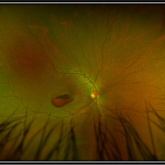
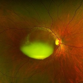
 Valsalva Retinopathy
Jul 30 2025 by Akansha Sharma
Color fundus photograph of a 33 year old male with subhyaloid hemorrhage suggestive of valsalva retinopathy.
Photographer: DR. AKANSHA SHARMA
Condition/keywords: subhyaloid hemorrhage, valsalva retinopathy
-
 Valsalva Retinopathy
Jul 30 2025 by Akansha Sharma
Color fundus photograph of a 33 year old male with subhyaloid hemorrhage suggestive of valsalva retinopathy.
Photographer: DR. AKANSHA SHARMA
Condition/keywords: subhyaloid hemorrhage, valsalva retinopathy
-
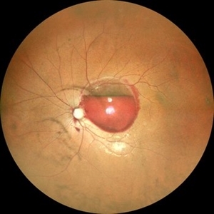
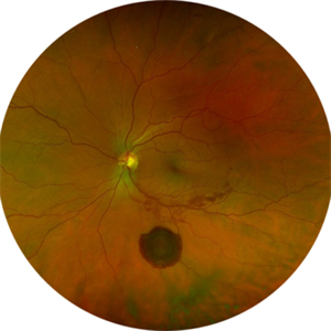
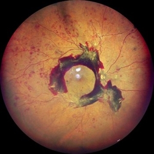
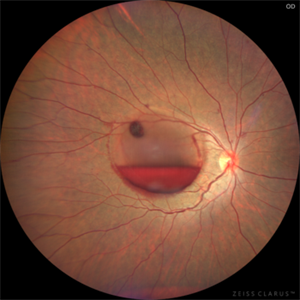
 Subhyaloid Hemorrhage
Jul 16 2025 by Paulina Araujo
55-degree central fundus photograph of the left eye (OS) shows a prominent subhyaloid hemorrhage in the inferior posterior pole, displaying a characteristic 'boat-shaped' appearance with well-defined margins and dark red coloration.
Photographer: Paulina D.Araujo Martínez, Asociación para Evitar la Ceguera en México I.A.P., Hospital Dr Luis Sánchez Bulnes.
Condition/keywords: subhyaloid hemorrhage
-
Subhyaloid Hemorrhage
Jul 14 2025 by SHRADDHA ASHOK CHANDORKAR, DNB DO FVRS
19 year old female presented with sudden blurring of vision in her right eye since few hours after she attended a DJ party the previous night. On examination Vision was counting fingers close to face and Retina showed Subhyaloid hemorrhage with some RPE damage. YAG hyaloidotomy was performed and the subhyaloid hemorrhage was drained. Need for injections if RPE damage and development of CNV in future was explained. Patient was apprehensive as the vision was not restored immediately after the blood was drained. On subsequent follow ups slowly patient’s vision was restored to 6/6N6 after about a month.
Condition/keywords: subhyaloid hemorrhage
-
 Subhyaloid Hemorrhage With Dispersed Vitreous Hemorrhage in a Case of Old Lasered Branch Retinal Vein Occlusion
Jul 12 2025 by Akansha Sharma
Color fundus photograph of a 32 year old hypertensive and diabetic male with subhyaloid hemorrhage with dispersed vitreous hemorrhage in a case of old lasered branch retinal vein occlusion.
Photographer: DR. AKANSHA SHARMA
Condition/keywords: branch retinal vein occlusion (BRVO), laser photocoagulation, SHH, subhyaloid hemorrhage, VH, vitreous hemorrhage
-
 Subhyaloid Hemorrhage
Jul 12 2025 by SHRADDHA ASHOK CHANDORKAR, DNB DO FVRS
19 year old female presented with sudden blurring of vision in her right eye since few hours after she attended a DJ party the previous night. On examination Vision was counting fingers close to face and Retina showed Subhyaloid hemorrhage with some RPE damage. YAG hyaloidotomy was performed and the subhyaloid hemorrhage was drained. Need for injections if RPE damage and development of CNV in future was explained. Patient was apprehensive as the vision was not restored immediately after the blood was drained. On subsequent follow ups slowly patient’s vision was restored to 6/6N6 after about a month.
Photographer: Dr.Shraddha Chandorkar
Imaging device: Zeiss
Condition/keywords: subhyaloid hemorrhage
-
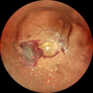
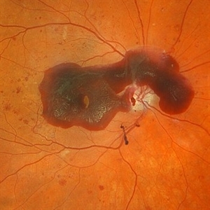
 Large Subhyaloid Hemorrhage
Jul 11 2025 by Jessilla Phou
This is a fundus photograph depicting a large subhyaloid hemorrhage in the mid periphery of the left eye. The patient, a 53-year-old female, presented with a sudden onset of floaters, headache, and blurred vision. The image also demonstrates associated optic disc hemorrhage, vitreous hemorrhage, retinal hemorrhage, and venous tortuosity. Despite the extensive workup performed and the severity of the hemorrhage, no underlying cause was determined.
Photographer: Jessilla Phou
Imaging device: Optos California
Condition/keywords: fundus photograph, optic disc hemorrhage, retinal hemorrhage, venous tortuosity, vitreous hemorrhage
-
 Chronic Sub-Hyaloid Hemorrhage with Dehemoglobinized Blood
Jul 11 2025 by Aditya S Kelkar, MS, FRCS, FASRS,FRCOphth
Fundus photograph of an 38-year-old man with a long standing sub hyaloid hemorrhage with dehemoglobinized blood.
Photographer: Optom Salomi Sonawane, National Institute of Ophthalmology, Pune, India
Imaging device: Optos Daytona
Condition/keywords: chronic, dehemoglobinized hemorrhage, SUBHYALOID HEMORRHAGE
-
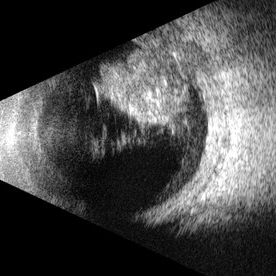 Glass
Glass
May 21 2025 by Gustavo Uriel Fonseca Aguirre
This B-mode transverse ultrasound scan reveals an intraocular glass foreign body secondary to penetrating trauma, with associated vitreous and subhyaloid hemorrhage. The glass fragments appear as hyperechoic linear structures in both the vitreous cavity and the retinachoroidal complex.
Photographer: Gustavo U. Fonseca Aguirre, Hospital Conde de Valenciana, Ciudad de México
Condition/keywords: glass, intraocular foreign body
-
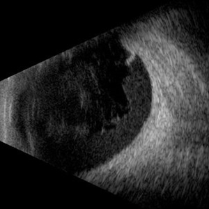 Hemorrhagic Vitreous Detachment
May 21 2025 by Gustavo Uriel Fonseca Aguirre
This B-mode longitudinal ultrasound scan shows a hemorrhagic vitreous detachment with the peripheral hyaloid strongly adherent to a retinal break. Associated vitreous and subhyaloid hemorrhage are present, indicating acute vitreoretinal traction.
Photographer: Gustavo U. Fonseca Aguirre, Hospital Conde de Valenciana, Ciudad de México
Condition/keywords: Hemorrhagic Vitreous Detachment
-
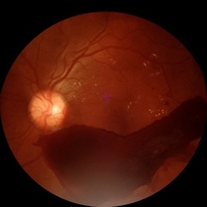
 High Risk Proliferative Diabetic Retinopathy with Sub-hyaloid Hemorrhage
May 13 2025 by Anupama Kiran Kumar
This image shows a case of high risk proliferative diabetic retinopathy. The retina is unlasered with a taut posterior hyaloid and a sub-hyaloid hemorrhage at the macula and along the arcades ,sparing the fovea.
Photographer: Mr Pratap
Imaging device: Mirante SLO/OCT (Nidek Co., Gamagori, Japan)
Condition/keywords: Diabetes, Diabetic Retinopathy, proliferative diabetic retinopathy (PDR), subhyaloid hemorrhage
-
Posterior Hyphema
Apr 29 2025 by Gustavo Uriel Fonseca Aguirre
This kinetic B-mode ultrasound scan (inferior transverse view) reveals combined vitreous and subhyaloid hemorrhage, accompanied by a mobile posterior hyphema level. The dynamic evaluation shows dependent blood shifting with positional changes, confirming fresh hemorrhage without organization.
Condition/keywords: diabetic retinopathy
-
 Advanced Proliferative Diabetic Retinopathy
Apr 9 2025 by Gustavo Uriel Fonseca Aguirre
B-mode ultrasound of a patient with long-standing poorly controlled diabetes demonstrates characteristic findings of advanced proliferative diabetic retinopathy. The examination reveals moderate vitreous hemorrhage appearing as diffuse hyperechoic opacities throughout the vitreous cavity, along with a posterior hyaloid membrane densely infiltrated by hemorrhagic material, showing irregular thickening and increased reflectivity. A mild subhyaloid hemorrhage is visible as a subtle hyphema-like space anterior to the retinal surface. The study documents a total tractional retinal detachment, evidenced by rigid retinal folds with clear insertion points of vitreous strands, accompanied by a significant subretinal hemorrhage seen as a prominent hyperechoic collection beneath the elevated retina. These findings collectively illustrate the severe vitreoretinal interface pathology characteristic of end-stage diabetic eye disease, with predominant tractional components and distinct echographic stratification of hemorrhagic layers - from anterior vitreous involvement to deeper subretinal blood accumulation.
Photographer: Gustavo U. Fonseca Aguirre, Hospital Conde de Valenciana, Ciudad de México
Condition/keywords: diabetic retinopathy, tractional retinal detachment, Vitreous hemorrhage
-
 Severe NPDR with Subhyaloid Hemorrhage
Apr 9 2025 by Kimberly Wakester
Optomap RGB of an 47 year-old man with severe NPDR with subhyaloid hemorrhage in the right eye.
Photographer: Kimberly Wakester, COA, OCT-C
Imaging device: Optos California
Condition/keywords: severe NPDR, subhyaloid hemorrhage
-
 The Pouring RAM
Mar 25 2025 by Shrishti mishra
A 63 year old male with RAM lesion in the right eye associated with multilayered hemorrhage.
Imaging device: Optos nikon
Condition/keywords: FFA, retinal arterial macroaneurysm, subhyaloid hemorrhage
-
 Hyaloid Butterfly
Mar 13 2025 by Gustavo Uriel Fonseca Aguirre
Axial ultrasound showing a phakic eye with vitreous hemorrhage, hyaloids impregnated with blood, hyalochisis (butterfly-shaped), subhyaloid hemorrhage, and retinal tractions involving the macular area.
Photographer: Gustavo U. Fonseca Aguirre, Hospital Conde de Valenciana, Ciudad de México
Condition/keywords: Hyaloschisis, Sub hyaloid haemorrhage, Vitreous hemorrhage
-
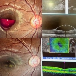
 Subhyaloid Hemorrhage
Mar 1 2025 by Vishal Agrawal, MD, FRCS,FACS,FASRS
A 37-year-old male presented with sudden diminution of vision in the right eye. On fundus examination boat shaped sub hyaloid hemorrhage was noted and a YAG hyaloidotomy was performed.
Photographer: Dr Ayushi Gupta
Imaging device: Clarus 700
Condition/keywords: Sub hyaloid haemorrhage, YAG HYALOIDOTOMY
-
 PDR
PDR
Feb 25 2025 by Parnian Arjmand, MD, MSc, FRCSC, DABO
Young patient with proliferative diabetic retinopathy and subhyaloid hemorrhage. Extensive neovascular fronds can be noted throughout the posterior pole and disc.
Condition/keywords: subhyaloid hemorrhage
-
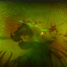
 Eales Disease
Jan 31 2025 by Thirumalesh Mochi Basavaraj, MD
Ultra wide field image of a 24 year-old young healthy adult male with a visible sea fan neovascularization with partial PVD secondary to Scatter LASER photocoagulation with Vitreous and subhyaloid hemorrhage.
Photographer: Puttaswamy N K
Condition/keywords: Eales disease, Neovascularisation elsewhere (NVE), sea fan
-
 Eales Disease
Jan 31 2025 by Thirumalesh Mochi Basavaraj, MD
Ultra-wide field image of a 24 year old young healthy adult male with a visible sea fan neovascularization with partial PVD with vitreous and subhyaloid hemorrhage.
Photographer: Puttaswamy
Condition/keywords: Eales disease, sea fan, Ultra-wide field retinal imaging
 Loading…