Search results (134 results)
-
 B-scan Ultrasound of Choroidal Melanoma with Serous Retinal Detachment
Sep 5 2025 by Kristen Wagner
B-scan ultrasound of a choriodal melanoma with serous retinal detachment.
Photographer: Kristen Wagner, COT Tennessee Retina
Condition/keywords: B scan ultrasound, Choroidal melanoma, serous retinal detachment
-
 Vogt-Koyanagi-Harada (VKH) Syndrome
Aug 18 2025 by Ricardo Leitão Guerra
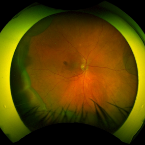
A 45 year old female presenting serous retinal detachment due to Vogt-Koyanagi-Harada (VKH) Syndrome
Photographer: Ricardo Leitão Guerra, Leitão Guerra - Oftalmologia
Imaging device: Zeiss Clarus 700
Condition/keywords: Vogt-Koyanagi-Harada (VKH) Symdrome
-
 Retinal Vasoproliferative Tumor
Jun 24 2025 by Marcelo Zas, MD PhD
We present a case of a 33-year-old male patient, who presented with decreased visual acuity in his right eye with 20/80, presenting a primary retinal vasoproliferative tumor in the lower temporal quadrant. The tumor is associated with serous retinal detachment, hard exudation, neovascularization and telangiectasias. Lipid exudates extend toward the macula, indicating macular involvement, which may contribute to decreased visual acuity. Oi was normal with 20/20 of BCVA. The patient was treated initially with IV anti-VEGF therapy and cryotherapy.
Photographer: Marcelo Zas MD PhD
Condition/keywords: RETINAL VASOPROLIFERATIVE TUMOR
-
 Serpiginous Choroidopathy
Jun 23 2025 by César Adrián Gómez Valdivia, MD
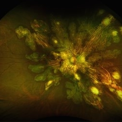
Fundus photograph of a 29 year-old female patient diagnosed with Serpiginous Choroidopathy. Finings were bilateral. The most common complication of SC is choroidal neovascularization affecting up to 35% of patients. Other reported complications are subretinal fibrosis, cystoid macular edema, branch vein occlusion, serous retinal detachment, optic disc neovascularization ,and anterior uveitis.
Photographer: @eyemissu2
Imaging device: TOPCON TRC-50DX
Condition/keywords: serpiginous choroiditis
-
 Serpiginous Choroidopathy
Jun 23 2025 by César Adrián Gómez Valdivia, MD
Fundus photograph of a 29 year-old female patient diagnosed with Serpiginous Choroidopathy. Finings were bilateral. The most common complication of SC is choroidal neovascularization affecting up to 35% of patients. Other reported complications are subretinal fibrosis, cystoid macular edema, branch vein occlusion, serous retinal detachment, optic disc neovascularization, and anterior uveitis.
Photographer: @eyemissu2
Imaging device: California ICG OPTOS
Condition/keywords: serpiginous choroiditis
-
 Choroidal Hemangioma
Jun 18 2025 by Moazzam Parvez
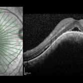
An OCT image of a 42 year old man presenting with a vision of 20/80 and complaining of distortion. OCT reveals serous retinal detachment with RPE alteration and disruption of outer retinal layers.
Photographer: Moazzam Parvez , Netralayam , Kolkata
Imaging device: Heidelberg Spectralis
Condition/keywords: Choroidal Hemangioma, Sub retinal fluid, tumor
-
 Giant RPE Rip with Serous Retinal Detachment
Apr 30 2025 by Amber Dubey
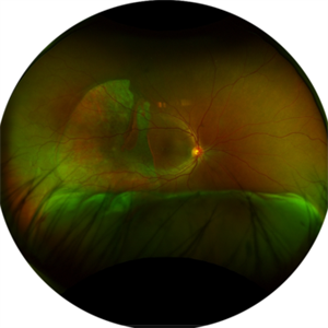
A 50 year-old man with sudden onset diminution of vision since 3 days. Optos Image showing a fovea-sparing giant RPE rip temporally with associated serous retinal detachment inferiorly without ocular and systemic co-morbidity or antecedent history of trauma.
Photographer: Dr. Amber Dubey, Sri Sankaradeva Nethralaya, Guwahati, India
Imaging device: Optos imaging system
Condition/keywords: Optos, RPE Rip, serous retinal detachment
-
 Posterior Nodular Scleritis
Apr 23 2025 by Gustavo Uriel Fonseca Aguirre
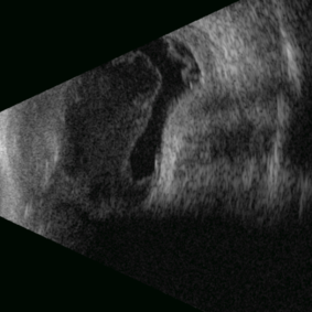
This B-mode ultrasound scan demonstrates a posterior scleral nodule accompanied by vitritis, serous retinal detachment, and annular choroidal detachment. The nodule appears as a localized hypoechoic scleral thickening, while the serous retinal detachment shows a smooth convex configuration. The choroidal detachment presents with the characteristic ring-shaped elevation, suggesting significant intraocular inflammation or hypotony.
Photographer: Gustavo U. Fonseca Aguirre, Hospital Conde de Valenciana, Ciudad de México
Condition/keywords: posterior nodular scleritis, posterior scleritis
-
 Serous Choroidal Detachment
Apr 18 2025 by Tracy Vu
Fundus photograph of a 77-year-old male with near 360 degree choroidal effusions accompanied by a mild serous choroidal detachment in the inferotemporal quadrant.
Condition/keywords: choroidal effusion, serous choroidal detachment, serous retinal detachment
-
 Choroidal Melanoma with Serous Retinal Detachment
Dec 20 2024 by Daniel Davis, OCT-C
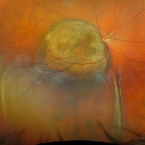
67 year old male presenting with large pigmented choroidal mass with serous retinal detachment.
Photographer: Daniel Davis, OCT-C, The Retina Institute
Imaging device: Optos California
Condition/keywords: Retina detachment
-
 Serous Retinal Detachment
Oct 14 2024 by César Adrián Gómez Valdivia, MD
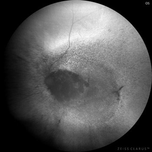
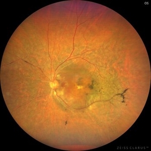
Serous Retinal Detachment found in a 19 year-old female patient with suspected Vogt-Koyanagi-Harada disease. Findings were bilateral. Patient was admitted for Methylprednisolone and Cyclophosphamide treatment.
Photographer: @eyemissu2
Imaging device: California ICG OPTOS
Condition/keywords: serous retinal detachment, vkh, Vogt-Koyanagi-Harada
-
 Serous Retinal Detachment
Oct 14 2024 by César Adrián Gómez Valdivia, MD
Serous Retinal Detachment found in a 19 year-old female patient with suspected Vogt-Koyanagi-Harada disease. Findings were bilateral. Patient was admitted for Methylprednisolone and Cyclophosphamide treatment.
Photographer: @eyemissu2
Imaging device: California ICG OPTOS
Condition/keywords: serous retinal detachment, Vogt-Koyanagi-Harada
-
 Morning glory disc anomaly-associated maculopathy: fibroglial tissue with a Mac-Off serous retinal detachment.
Jun 26 2024 by Julián Villarreal, MD
19 year old with a Morning glory disc anomaly-associated maculopathy: fibroglial tissue with a Mac-Off serous retinal detachment.
Photographer: Julián Villarreal MD
Imaging device: Mirante
Condition/keywords: fibroglial tissue, Morning Glory Anomaly, retinal detachment of the macula
-
 Serous Retinal Detachment in Advanced Proliferative Diabetic Retinopathy
Feb 15 2024 by Annaka Gooding
Ultra-Wide fundus photograph of a 29 year old female with a Serous Retinal Detachment in Advanced PDR. Patient present to clinic with LP vision following PPV and fill in PRP. Physician recommended oral prednisone treatment and to reassess at their following visit.
Photographer: Annaka Gooding, CPO
Imaging device: Optos California RGB
Condition/keywords: Diabetes, diabetic macular edema, fundus photography, OPTOS CALIFORNIA, pan-retinal photocoagulation (PRP), pars plana vitrectomy (PPV), proliferative diabetic retinopathy (PDR), serous retinal detachment, ultra-wide field imaging
-
 Choroidal Melanoma
Sep 7 2023 by Annaka Gooding
Ultra-Widefield pseudo-color and autofluorescence imaging of a 59 year old male with Choroidal Melanoma affecting his left eye. Patient reported floaters OS for months prior to examination as well as 1-2 weeks of "tunnel vision". Patient denies personal history of cancer. Patient's vision at time of examination was CF@5FT. Due to the Tumor size, the patient has developed a serous retina detachment in their inferior retina
Photographer: Annaka Gooding
Imaging device: Optos California
Condition/keywords: autofluorescence imaging, choroidal tumor, fundus photography, OPTOS CALIFORNIA, serous retinal detachment
-
 VKH - Uveitic stage
Jun 23 2023 by Sergio Emilio Sifuentes Renteria, MD
Fundus photograph of a young female with VKH in uveitic stage
Photographer: Sergio Emilio Sifuentes Rentería - Foundation Hospital Nuestra Señora de La Luz
Condition/keywords: choroiditis, panuveitis, serous retinal detachment, VKH, Vogt-Koyanagi-Harada
-
 acute posterior multifocal placoid pigment epitheliopathy
Sep 23 2022 by Jaideep sharma
A 50-year old woman presented to us with unilateral progressive and painless visual blurring. She was diagnosed as a case of CSCR and started on topical dorzolamide with no improvement in VA. Her best-corrected visual acuity (BCVA) was RE 6/6 and LE 6/60 . Eye examination revealed vitritis (grade1) with optic disc hyperemia and multiple serous retinal detachments with choroidal striae in the left eye and a normal right eye. She is k/c/o diabetes. Her past ocular and drug histories were unremarkable. Retinal imaging revealed characteristic features of APMPPE in the left eye. All laboratory testing results were inconclusive. VA and OCT findings significantly improved following the treatment with LE posterior sub tenon’s triamcinolone (40 mg/ml). 1 month post injection VA of the left eye reached 6/6 with resolved serous retinal detachments in this eye. This case is unique as it was managed via PST injection rather than conventional steroid therapy
Photographer: jaideep sharma jaipur calgary eye hospital rajasthan india
Condition/keywords: acute posterior multifocal placoid pigment epitheliopathy (APMPPE), FFA
-
 Acute posterior multifocal placoid pigment epitheliopathy (APMPPE)
Sep 23 2022 by Jaideep sharma
A 50-year old woman presented to us with unilateral progressive and painless visual blurring. She was diagnosed as a case of CSCR and started on topical dorzolamide with no improvement in VA. Her best-corrected visual acuity (BCVA) was RE 6/6 and LE 6/60 . Eye examination revealed vitritis (grade1) with optic disc hyperemia and multiple serous retinal detachments with choroidal striae in the left eye and a normal right eye. She is k/c/o diabetes. Her past ocular and drug histories were unremarkable. Retinal imaging revealed characteristic features of APMPPE in the left eye. All laboratory testing results were inconclusive. VA and OCT findings significantly improved following the treatment with LE posterior sub tenon’s triamcinolone (40 mg/ml). 1 month post injection VA of the left eye reached 6/6 with resolved serous retinal detachments in this eye. This case is unique as it was managed via PST injection rather than conventional steroid therapy
Photographer: jaideep sharma jaipur calgary eye hospital rajasthan india
Condition/keywords: acute posterior multifocal placoid pigment epitheliopathy (APMPPE), FFA
-
 Choroid hemangioma
Sep 7 2022 by JEFFERSON R SOUSA, Tecg.º (Biomedical Systems Technology)
Patient 54 years old, Female, progressive loss of vision. In the multimodal evaluation of the retina showed important retinal alterations. A discreet opacity of the media impairs the quality of the images. In the Autofluorescent Background Image with a green filter, because it reaches a depth in the retinal tissue, it is able to show changes that affect the retinal pigment epithelium, it was better in this case than with the green filter. WF retinography shows an elevated, slightly reddish lesion, probable serous retinal detachment, mobilization of pigments and phantom vessels.
Photographer: JEFFERSON ROCHA DE SOUSA - Retinal Department at Instituto Dr. Suel Abujamra Sao Paulo-Brazil
Imaging device: Clarus 700 - Zeiss 135 degree images. Multimodal Evaluation
Condition/keywords: elevated retinal lesion, hemangioma, melanoma, serous retinal detachment
-
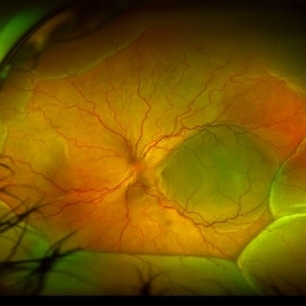 Serous Retinal Detachment in a patient with Vogt-Koyanagi-Harada Syndrome
Aug 24 2021 by Nicolás Crim, MD
Fundus OS. Female 35 year-old with bilateral Serous Retinal Detachment with acute lost of visual acuity.
Photographer: Nicolas Crim MD, Córdoba, Argentina
Condition/keywords: Vogt-Koyanagi-Harada
-
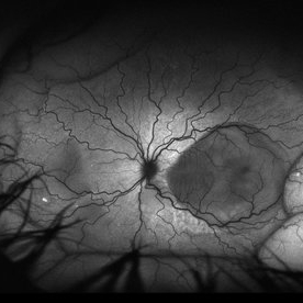 Serous Retinal Detachment in a patient with Vogt-Koyanagi-Harada syndrome
Aug 24 2021 by Nicolás Crim, MD
Autofluorescence OS. 35 year-old female with bilateral serous retinal detachment with acute loss of visual acuity.
Photographer: Nicolas Crim MD, Córdoba, Argentina
Condition/keywords: serous retinal detachment, vision loss, Vogt-Koyanagi-Harada
-
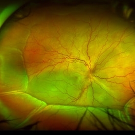 Serous Retinal Detachment in a patient with Vogt-Koyanagi-Harada syndrome
Aug 24 2021 by Nicolás Crim, MD
Fundus OD. 35 year-old female with bilateral serous retinal detachment with acute loss of visual acuity
Photographer: Nicolas Crim MD, Córdoba, Argentina
Condition/keywords: serous retinal detachment, vision loss, Vogt-Koyanagi-Harada
-
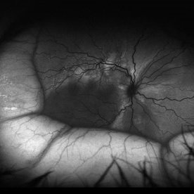 Serous Retinal Detachment in a patient with Vogt-Koyanagi-Harada Syndrome
Aug 24 2021 by Nicolás Crim, MD
Autofluorescence OD. 35 year-old female with bilateral serous retinal detachment with acute loss of visual acuity.
Photographer: Nicolas Crim MD, Córdoba, Argentina
Condition/keywords: Vogt-Koyanagi-Harada
-
 Ciliochoroidal Melanoma with Total Serous Retinal Detachment
Jun 20 2021 by Jesus Lozano, MD
64-year-old woman with a ciliochoroidal melanoma after SRS with a exudative retinal detachment.
Photographer: Dr. Jesús Lozano Gutiérrez. Hadassah Medical Center, Israel.
Imaging device: Slit lamp BI 900 camera
Condition/keywords: cataract, exudative detachment
 Loading…