Search results (98 results)
-
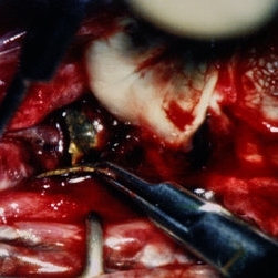
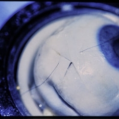
 Intraocular Foreign Body Scleral Lac
Nov 19 2025 by Nikhil Das, M.D.
A 34-year-old man presented with a right intraocular foreign body after hammering a carbon-steel chisel 12 hours after injury. CT orbits showed a 3-mm hyperattenuating foreign body within the right globe, centered in the vitreous cavity. BCVA was 20/40. Anterior segment examination revealed a 2.8-mm scleral laceration. DFE demonstrated a metallic IOFB, a superior air bubble, superior commotio retinae, and Berlin’s edema involving the macula.
Photographer: Nikhil Das, Saint Louis University School of Medicine
Imaging device: iPhone
Condition/keywords: intraocular foreign body, iofb, metallic foreign body, scleral laceration
-
 Intraocular Foreign Body CT Coronal
Nov 19 2025 by Nikhil Das, M.D.
A 34-year-old man presented with a right intraocular foreign body after hammering a carbon-steel chisel 12 hours after injury. CT orbits showed a 3-mm hyperattenuating foreign body within the right globe, centered in the vitreous cavity. BCVA was 20/40. Anterior segment examination revealed a 2.8-mm scleral laceration. DFE demonstrated a metallic IOFB, a superior air bubble, superior commotio retinae, and Berlin’s edema involving the macula.
Photographer: Nikhil Das, Saint Louis University School of Medicine
Imaging device: CT Scan
Condition/keywords: intraocular foreign body, iofb, metallic foreign body, scleral laceration
-
 Intraocular Foreign Body CT Axial
Nov 19 2025 by Nikhil Das, M.D.
A 34-year-old man presented with a right intraocular foreign body after hammering a carbon-steel chisel 12 hours after injury. CT orbits showed a 3-mm hyperattenuating foreign body within the right globe, centered in the vitreous cavity. BCVA was 20/40. Anterior segment examination revealed a 2.8-mm scleral laceration. DFE demonstrated a metallic IOFB, a superior air bubble, superior commotio retinae, and Berlin’s edema involving the macula.
Photographer: Nikhil Das, Saint Louis University School of Medicine
Imaging device: CT Scan
Condition/keywords: intraocular foreign body, iofb, metallic foreign body, scleral laceration
-
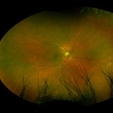
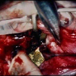
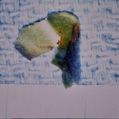
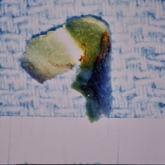
 Intraocular Foreign Body
Nov 19 2025 by Nikhil Das, M.D.
A 34-year-old man presented with a right intraocular foreign body after hammering a carbon-steel chisel 12 hours after injury. CT orbits showed a 3-mm hyperattenuating foreign body within the right globe, centered in the vitreous cavity. BCVA was 20/40. Anterior segment examination revealed a 2.8-mm scleral laceration. DFE demonstrated a metallic IOFB, a superior air bubble, superior commotio retinae, and Berlin’s edema involving the macula.
Photographer: Nikhil Das, Saint Louis University School of Medicine
Condition/keywords: intraocular foreign body, metallic foreign body, scleral laceration
-
 Posterior Scleral Laceration
May 24 2022 by Ahmad B. Tarabishy, MD
A 49 year old male was referred from the ER following an injury to his right medial eyelid with a sharp metal tip. He had brief pain at the time. No new floaters, flashes, or blurred vision. Intraocular pressure was 18 OS. Examination showed a full thickness laceration of the nasal posterior globe with adjacent hemorrhage. Prophylactic laser coagulation was performed. Examination 2 weeks later shows maturing laser scars and no complications related to the scleral laceration. The patient reports no new vision changes.
Photographer: Angelo Rico MD, Retina Specialists of Tampa
Imaging device: Optos
Condition/keywords: corneal laceration, globe perforation
-
 Posterior Scleral Laceration
May 24 2022 by Ahmad B. Tarabishy, MD
A 49 year old male was referred from the ER following an injury to his right medial eyelid with a sharp metal tip. He had brief pain at the time. No new floaters, flashes, or blurred vision. Intraocular pressure was 18 OS. Examination showed a full thickness laceration of the nasal posterior globe with adjacent hemorrhage. Prophylactic laser coagulation was performed. Examination 2 weeks later shows maturing laser scars and no complications related to the scleral laceration. The patient reports no new vision changes.
Photographer: Angelo Rico MD, Retina Specialists of Tampa
Imaging device: Optos
Condition/keywords: globe perforation, scleral laceration
-
 Scleral Laceration from Metallic Foreign Body
Feb 18 2022 by Maxwell J Wingelaar, MD
A young male presented with irritation after sustaining a metallic foreign body injury to his eye.
Photographer: Jarrod Wehmeier
Condition/keywords: Trauma
-
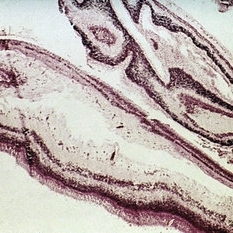 Slide 7-89
Feb 25 2019 by Lancaster Course in Ophthalmology
Strips of retina removed from a scleral laceration.
Condition/keywords: scleral laceration
-
 Intraocular Eyelash
Jan 2 2019 by John S. King, MD
34-year-old white male injured while taking apart wooden pallets with a hammer in each hand and no protective eye-wear; he did not notice what hit his eye; just said his eye hurt and teared for four days before calling an eye doctor; his vision was 20/400 sc and IOP 6; the anterior chamber was deep with minimal cell and no hypopyon; and conjunctival/scleral laceration was present near the lateral rectus insersion; the vitreous was quiet; in the temporal portion of the fundus and full thickness laceration was seen with surrounding hemorrhage and what appeared to be an eyelash vs other (possibly a staple, from the wooden pallet). During surgery 2 eyelashes were pulled from the area of the laceration; the lateral rectus muscle was disorganized; after primary closure, a ppv was performed, the object in the picture was removed with forcepts; laser partial afx and gas; antibiotics injected at 1/2 dose.
Photographer: Maisee Yang
Imaging device: Optos CA
Condition/keywords: intraocular foreign body, ruptured globe
-
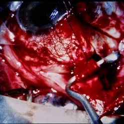
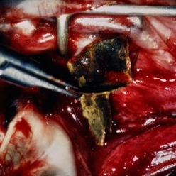 Trauma – Surgical View – Long Scleral Laceration and Foreign Body
Aug 11 2015 by H. Michael Lambert, MD
Further view of same eye, posterior laceration and large metallic foreign body.
Condition/keywords: intraocular foreign body, trauma
-
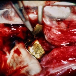 Trauma – Surgical View – Long Scleral Laceration and Foreign Body
Aug 11 2015 by H. Michael Lambert, MD
Further view of same eye, posterior laceration and large metallic foreign body.
Condition/keywords: intraocular foreign body, trauma
-
 Trauma – Surgical View – Long Scleral Laceration and Foreign Body
Aug 11 2015 by H. Michael Lambert, MD
Further view of same eye, posterior laceration and large metallic foreign body.
Condition/keywords: intraocular foreign body, trauma
-
 Trauma – Surgical View – Long Scleral Laceration and Foreign Body
Aug 11 2015 by H. Michael Lambert, MD
Further view of same eye, posterior laceration and large metallic foreign body.
Condition/keywords: intraocular foreign body, trauma
-
 Trauma – Surgical View – Long Scleral Laceration and Foreign Body
Aug 11 2015 by H. Michael Lambert, MD
Further view of same eye, posterior laceration and large metallic foreign body.
Condition/keywords: intraocular foreign body, trauma
-
 Trauma – Surgical View – Long Scleral Laceration and Foreign Body
Aug 11 2015 by H. Michael Lambert, MD
Further view of same eye, posterior laceration and large metallic foreign body.
Condition/keywords: intraocular foreign body, trauma
-
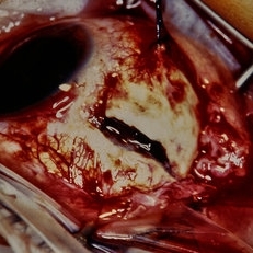 Trauma – Posterior Scleral Laceration
Aug 11 2015 by H. Michael Lambert, MD
Trauma – posterior scleral laceration.
Condition/keywords: scleral laceration, trauma
-
 Trauma – Posterior Scleral Laceration
Aug 11 2015 by H. Michael Lambert, MD
Posterior scleral laceration – before conjunctiva lifted.
Condition/keywords: scleral laceration, trauma
-
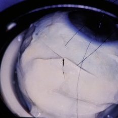
 Trauma – Eye Bank Eye With Stellate Scleral Laceration
Aug 11 2015 by H. Michael Lambert, MD
Showing repair of scleral laceration.
Condition/keywords: scleral laceration, trauma
-
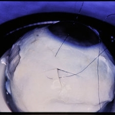
 Trauma – Eye Bank Eye With Stellate Scleral Laceration
Aug 11 2015 by H. Michael Lambert, MD
Showing repair of scleral laceration.
Condition/keywords: scleral laceration, trauma
-
 Trauma – Eye Bank Eye With Stellate Scleral Laceration
Aug 11 2015 by H. Michael Lambert, MD
Showing repair of scleral laceration.
Condition/keywords: scleral laceration, trauma
-
 Trauma – Eye Bank Eye With Stellate Scleral Laceration
Aug 11 2015 by H. Michael Lambert, MD
Showing repair of scleral laceration.
Condition/keywords: scleral laceration, trauma
 Loading…