Search results (10 results)
-
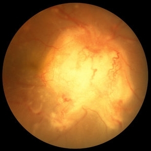
 Multimodal Imaging for Differentiating Unilateral Pseudo Optic Disc Swelling(Buried Drusen) From True Optic Disc Swelling
Feb 7 2024 by Fawwaz F Al Mamoori, MD, Medical Retina Consultant
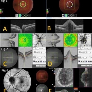
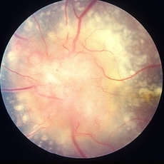
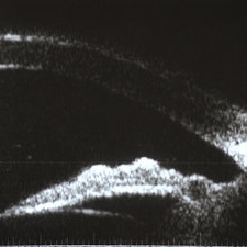
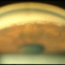
27-year-old male, medically free, presented with left unilateral optic disc swelling. BCVA=1.0(OU), color vision, and contrast sensitivity were normal (OU)with no RAPD in the left eye. Swept Source OCT: showed elevated left optic disc with hyporeflective mass (Fig-1 B). Enface OCT: Showed left peripapillary multiple ovoid mass lesions(drusen) (Fig-2 d, Fig3 F). FAF: of the left eye showed superonasal hyper autofluorescent drusenoid lesions)(Fig3 E). Orbital MRI with contrast was requested to exclude any compressive lesions like tumors(menigioma)or inflammatory lesions like granuloma(sarcoid granuloma). orbital MRI result was normal.
Photographer: Hana.S.Owais
Imaging device: TRITON(TOPCON,Swept Source OCT)
Condition/keywords: fundus autofluorescence (FAF), multimodal imaging, OCT EN FACE, optic disc drusen, optic disc edema, swept source
-
 Sarcoidosis
Mar 27 2023 by Joshua Friedman
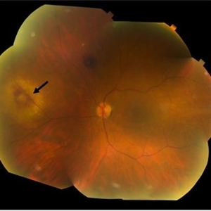
Fundus photograph of a 70-year-old woman with presumed sarcoidosis and sarcoid-associated choroidal granuloma.
Photographer: Christopher D Conrady, MD, PhD
Condition/keywords: sarcoid granuloma
-
 Slide 9-31
Feb 26 2019 by Lancaster Course in Ophthalmology
Sarcoidosis with retinal and vitreous involvement. A Daten-Fuchs-like nodule of sarcoid granuloma is present (lower right).
Condition/keywords: Dalen-Fuchs nodules, sarcoid granuloma, sarcoidosis
-
 Bilateral Optic Nerve Involvement in Sarcoidosis
Feb 25 2013 by Henry J. Kaplan, MD
Optic nerve granuloma of sarcoidosis in the right eye of a patient with bilateral involvement #1. Left eye is in the following slide.
Condition/keywords: bilateral involvement, sarcoid granuloma
-
 Bilateral Optic Nerve Involvement in Sarcoidosis
Feb 25 2013 by Henry J. Kaplan, MD
Optic nerve head granuloma of sarcoidosis with severe infiltration and exudation in the left eye of the same patient #2.
Condition/keywords: bilateral involvement, sarcoid granuloma
-
 Sarcoid Sarcoidosis
Feb 13 2013 by From the Collections of Thomas M. Aaberg, MD and Thomas M. Aaberg Jr., MD
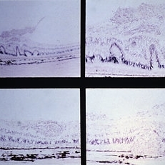
Drawing of retinal sarcoidosis, cultaneous sarcoid granuloma.
Condition/keywords: candle wax dripping, sarcoid granuloma, sarcoidosis, vasculitis
-
 Sarcoidosis Panuveitis Slide 4
Oct 22 2012 by Ronald C. Gentile, MD
High frequency ultrasound biomicroscopy of the anterior chamber and angle images a granuloma involving the iris root and Bussaca nodules on the iris surface consistent with granulomatous uveitis.
Photographer: The New York Eye & Ear Infirmary Department of Medical Imaging
Condition/keywords: sarcoid granuloma, sarcoidosis panuveitis
-
 Sarcoidosis Panuveitis Slide 3
Oct 22 2012 by Ronald C. Gentile, MD
Gonioscopic photograph reveals peripheral anterior synechiae with granuloma involving the iris root and Bussaca nodules on the iris surface consistent with granulomatous uveitis.
Photographer: The New York Eye & Ear Infirmary Department of Medical Imaging
Condition/keywords: sarcoid granuloma, sarcoidosis panuveitis
-
 Sarcoid Granuloma of Optic Nerve
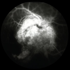
Oct 9 2012 by Jeffrey G. Gross, MD, FASRS
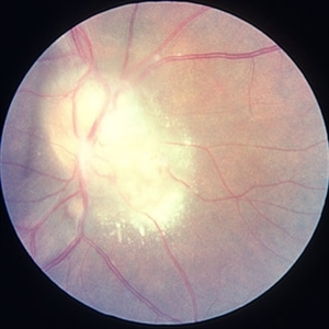
Sarcoid granuloma of optic nerve, FA, early phase, 20/400.
Condition/keywords: 20/400, autoimmunity, early phase, sarcoid granuloma, sarcoidosis
-
 Sarcoid Granuloma of Optic Nerve
Oct 9 2012 by Jeffrey G. Gross, MD, FASRS
Sarcoid granuloma of optic nerve, 20/400.
Condition/keywords: 20/400, autoimmunity, sarcoid granuloma, sarcoidosis
 Loading…