Search results (80 results)
-
 Retinopathy of Prematurity
Oct 26 2025 by Anjana Mirajkar, MS Ophthalmology
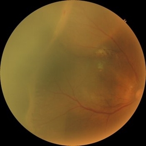
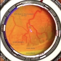
Fundus photograph of right eye of premature baby showing stage 3 in zone 2 posterior.
Photographer: Dr. Anjana Mirajkar- HV desai eye hospital ,Pune
Imaging device: Retcam
Condition/keywords: retinopathy of prematurity (ROP), stage 3
-
 Retinopathy of Prematurity
Oct 26 2025 by Anjana Mirajkar, MS Ophthalmology
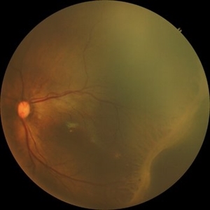
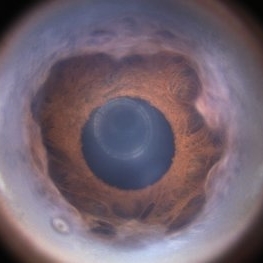
Fundus photograph of a left eye of a premature baby showing stage 3 in zone 2 posterior.
Photographer: Dr. Anjana Mirajkar- HV desai eye hospital ,Pune
Imaging device: Retcam
Condition/keywords: retinopathy of prematurity (ROP), retinopathy of prematurity stage 3
-
 Retinopathy of Prematurity
Oct 26 2025 by Anjana Mirajkar, MS Ophthalmology
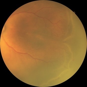
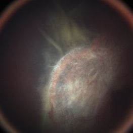
Fundus photograph of left eye premature baby having stage 3 in zone 2A with a secondary notch.
Photographer: Dr. Anjana Mirajkar- HV Desai eye hospital ,Pune
Imaging device: retcam
Condition/keywords: retinopathy of prematurity (ROP), stage 3
-
 Fluorescein Angiogram of ROP With Cryo Scarring
Jul 7 2025 by Jenn Geelan
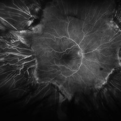
FA photo of a 34 year old male with prior stage 3 ROP with history of 360 degree cryotherapy.
Photographer: Jenn Geelan, Retina-Vitreous Surgeons of CNY
Imaging device: Optos California
Condition/keywords: cryotheraphy scar, fluorescein angiogram (FA), fundus photograph, retinopathy of prematurity (ROP), ROP, tilted disc
-
 Aggressive Posterior Retinopathy of Prematurity (APROP)
May 16 2025 by KANWALJEET HARJOT MADAN, M.S. (Ophthalmology); FAICO (Vitreous - Retina)
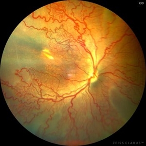
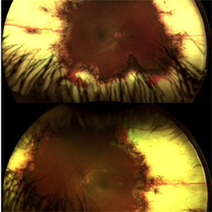
This is the fundus picture of right eye of a premature neonate depicting Aggressive Posterior Retinopathy of Prematurity (APROP). It is a severe rapidly progressing form of retinopathy that can lead to vision loss and blindness. It requires prompt diagnosis and treatment in the form of anti-VEGF agents and laser photocoagulation.
Photographer: Dr. Kanwaljeet Harjot Madan, Thind Eye Hospital, Jalandhar City (Punjab) INDIA.
Imaging device: Zeiss Clarus
Condition/keywords: Oxygen Exposure, retinopathy of prematurity (ROP)
-
 Comets in the Eye (Retinopathy of Prematurity)
Apr 8 2025 by KANWALJEET HARJOT MADAN, M.S. (Ophthalmology); FAICO (Vitreous - Retina)
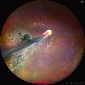
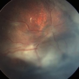
This is the fundus picture of right eye (RE) of a 4 years female child presented with outward deviation of right eye. Her parents also complained of diminution of vision in both eyes. On examination, her best corrected vision in RE was hand movements close to face and was 20/80 in LE. Posterior segment exam revealed presence of macular scar in RE and presence of dry retinal fold with dragging of retinal vessels. LE fundus revealed presence of nasal drag of optic disc. Parents gave history of untreated ROP as an infant. Retinopathy of Prematurity (ROP) is a Vaso proliferative disorder of Retina occurring in premature infants. Advances in neonatal care and ROP treatment has led these babies to live longer with this disease.
Photographer: Dr. Kanwaljeet Harjot Madan, Thind Eye Hospital, Jalandhar City (Punjab) INDIA.
Imaging device: Zeiss Fundus Camera
Condition/keywords: Retinopathy of Prematurity, Vaso proliferative disorder
-
 Comets in the Eye (Retinopathy of Prematurity)
Apr 8 2025 by KANWALJEET HARJOT MADAN, M.S. (Ophthalmology); FAICO (Vitreous - Retina)
This is the fundus picture of right eye (RE) of a 4 years female child presented with outward deviation of right eye. Her parents also complained of diminution of vision in both eyes. On examination, her best corrected vision in RE was hand movements close to face and was 20/80 in LE. Posterior segment exam revealed presence of macular scar in RE and presence of dry retinal fold with dragging of retinal vessels. LE fundus revealed presence of nasal drag of optic disc. Parents gave history of untreated ROP as an infant. Retinopathy of Prematurity (ROP) is a Vaso proliferative disorder of Retina occurring in premature infants. Advances in neonatal care and ROP treatment has led these babies to live longer with this disease.
Photographer: Dr. Kanwaljeet Harjot Madan, Thind Eye Hospital, Jalandhar City (Punjab) INDIA.
Imaging device: Zeiss Fundus Camera
Condition/keywords: Retinopathy of Prematurity
-
 Fight for Sight
Mar 26 2024 by Tushar Agrawal
Fundus photograph showing 28 weeker APROP; regressed well after ROP Laser photocoagulation as seen at age 3 months.
Imaging device: Retcam neo
Condition/keywords: aggressive posterior retinopathy of prematurity (APROP), pediatric retina, retinopathy of prematurity (ROP)
-
 Type-1 ROP treated with laser
Nov 25 2022 by Alexandre Grandinetti, MD, PhD
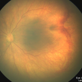
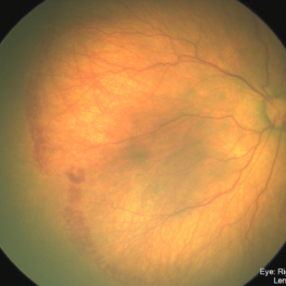
This picture shows the case of a 8-year-old girl born with 26 weeks of gestation, who was treated with laser photocoagulation due to type-1 ROP
Photographer: Corina Szrek, Hospital de Olhos do Paraná
Imaging device: California
Condition/keywords: retinopathy of prematurity (ROP), rop laser
-
 ROP-Zone-I-Stage-3-Plus
Jun 3 2022 by Dipak Nag, MBBS, FCPS, MSc, FRF
Fundus photograph of a child of gestational age 26 weeks and birth weight 1050 grams, shows dilatation and tortuosity of vessels in zone I, extra-retinal fibro-vascular proliferation, hemorrhage with huge peripheral avascular area.
Photographer: Dipak Nag, National Institute of Ophthalmology, Dhaka, Bangladesh
Imaging device: RetCam shuttle
Condition/keywords: retinopathy of prematurity (ROP), retinopathy of prematurity Plus disease, retinopathy of prematurity stage 3, retinopathy of prematurity zone I
-
 ROP 4B late Retinal Findings
Mar 31 2022 by Franco Benvenuto, MD
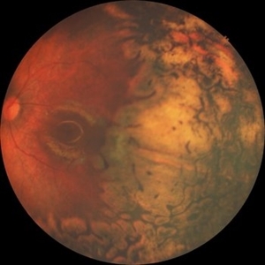
A 9-year-old male, that was born at 30 weeks of gestation with birth weight of 1500 g and history of hospitalization for 20 days with respiratory distress and packed red blood cell transfusion for anemia. At the first exam, both eyes were with stage 4B ROP. Vitrectomy with 25 G was done in both eyes. The flat fibrosis dragged the macula nasally in both the eyes.
Photographer: Franco Benvenuto, Universidad de Buenos Aires, Argentina; Universidad de Guadalajara, México.
Condition/keywords: cicatricial retinopathy of prematurity, retinopathy of prematurity (ROP)
-
 ROP 5A
Jan 24 2022 by Alexandre Grandinetti, MD, PhD
ROP retinal detachment.
Photographer: Alexandre Grandinetti
Imaging device: RetCam
Condition/keywords: retinopathy of prematurity (ROP), total retinal detachment
-
 Retinopathy of Prematurity
Aug 26 2021 by Stefanie Palmer
Patient has neovascular ridge temporal with elevation of vessels above the PRP scars. The image was obtained with the flying baby technique.
Photographer: Stefanie Palmer, CRA
Condition/keywords: retinopathy of prematurity (ROP)
-
 Retinopathy of Prematurity
Aug 26 2021 by Stefanie Palmer
Patient has neovascular ridge temporal with elevation of vessels above the PRP scars. The image was obtained with the flying baby technique.
Photographer: Stefanie Palmer, CRA
Condition/keywords: retinopathy of prematurity (ROP)
-
 Retinal Detachment post ROP
Jul 22 2021 by Vishal Gupta, MBBS, MS
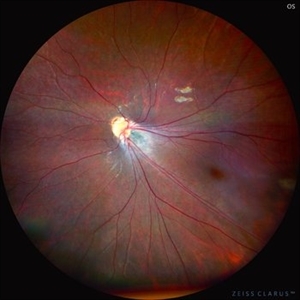
Fundus image of total Retinal Detachment in a five-year-old male kid with a history of prematurity.
Photographer: Dr Shobhit Chawla, Prakash Netra Kendr, Lucknow, UP, INDIA
Imaging device: Zeiss Clarus
Condition/keywords: retinopathy of prematurity (ROP)
-
 Retinopathy of Prematurity
Jul 12 2021 by Stefanie Palmer
Retinopathy of prematurity Stage 3 in a 5 month old baby. The flying baby technique was used to create this image.
Photographer: Stefanie Palmer CRA
Imaging device: scanning laser ophthalmoscope
Condition/keywords: retinopathy of prematurity (ROP), retinopathy of prematurity stage 3
-
 Fundus with ROP OS
Feb 4 2021 by Abdallah Mahmoud
Fundus with retinopathy of prematurity OS
Condition/keywords: fundus photograph, retinopathy of prematurity (ROP)
-
 Fundus with ROP OD
Feb 4 2021 by Abdallah Mahmoud
Fundus with retinopathy or prematurity OD.
Condition/keywords: fundus photograph, OD, retinopathy of prematurity (ROP)
-
 APROP 3-Month
Sep 29 2020 by Sham Talati, DOMS
A 3-month-old child of of APROP.
Photographer: Dr. Sham Talati,Retina Foundation,Ahmedabad
Imaging device: Nidek Mirante
Condition/keywords: aggressive posterior retinopathy of prematurity (APROP), retinopathy of prematurity (ROP)
-
 ROP
ROP
-
 Stage 1 ROP
Jun 4 2020 by Audina M. Berrocal, MD FASRS
Color photograph of the right eye in a premature baby. There is no plus disease, or evidence of Stage 1 in Zone 2 ROP. Stage 1 is defined as a line that separates vascularized retina to avascular retina.
Condition/keywords: retinopathy of prematurity (ROP), retinopathy of prematurity stage 1
-
 Retinopathy of Prematurity, Flying Baby Imaging Technique
May 27 2020 by Jamin S. Brown, MD
7-month-old male with improving ROP, was imaged using the flying baby technique and the use of a lid speculum.
Photographer: Stefanie Palmer, Retina-Vitreous Surgeons of CNY
Imaging device: California Optos
Condition/keywords: retinopathy of prematurity (ROP)
-
 Flying Baby Imaging Technique
May 27 2020 by Jamin S. Brown, MD
7-month-old male with improving ROP.
Photographer: Stefanie Palmer CRA, Retina-Vitreous Surgeons of CNY
Condition/keywords: retinopathy of prematurity (ROP)
-
 Retinopathy of Prematurity S/P Laser Complications
Apr 30 2020 by Giselle DeOliveira
Gonio photograph of 13-year-old female with retinopathy of prematurity, s/p laser complications.
Photographer: Giselle DeOliveira
Imaging device: Retcam III
Condition/keywords: laser, retinopathy of prematurity (ROP)
-
 Retinal Detachment
Apr 30 2020 by Giselle DeOliveira
Fundus Photograph of 13-month old male infant with retinopathy of prematurity, retinal detachment and Cohen Syndrome.
Photographer: Giselle DeOliveira, University of Miami, Bascom Palmer Eye Institute
Imaging device: Retcam III
Condition/keywords: retinopathy of prematurity (ROP)
 Loading…