Search results (389 results)
-
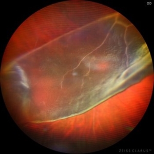
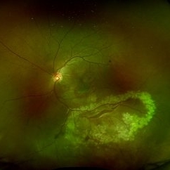
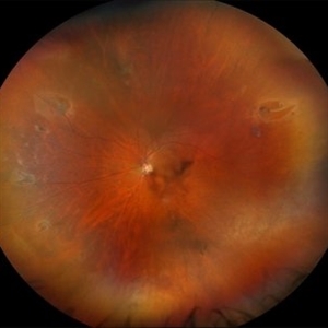
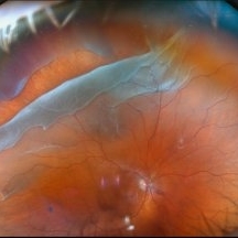
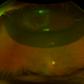
 Rhegmatogenous Retinal Detachment
Nov 27 2025 by Jacob Adrián Ruíz García
Ultra–widefield fundus image demonstrates an extensive rhegmatogenous retinal detachment (RRD) with several large, irregular retinal tears are visible in the temporal retina, with associated surrounding subretinal fluid. The detachment appears bullous, with fluid extending widely across the mid-periphery and involving much of the posterior pole.
Photographer: Jacob Adrián Ruíz Garcia, Fundación Hospital Nuestra Señora de la Luz I.A.P, México City
Imaging device: Optos California
Condition/keywords: horseshoe tear, Rhegmatogenous retinal detachment, tear
-
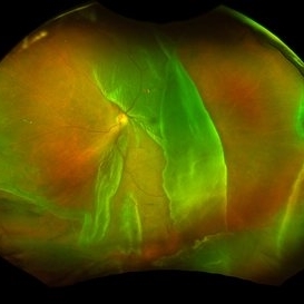
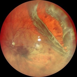
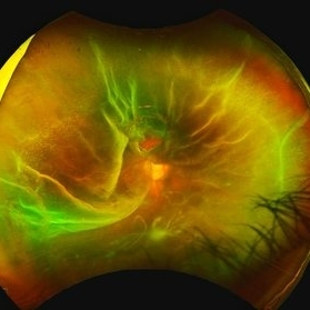
 Bullous Retinal Detachment
Nov 13 2025 by Virginia Gebhart
42 year old female referred for vision loss x 4-5 days. Bullous retinal detachment from 8:00 to 3:00 with retinal tear at 11:00. Macula is detached. Vision is LP, IOP of 3. Pt is scheduled for GFE and possible scleral buckle.
Photographer: Virginia Gebhart, Retina Consultants of Carolina
Imaging device: Optos California
Condition/keywords: bullous retinal detachment, retinal detachment, retinal detachment of the macula, retinal tear with detachment
-
 A Dramatic Curtain Call in the Retina
Oct 13 2025 by Tejaswita Verma
Fundus image of a 15 year old boy with 3 day history of sudden DOV, IOP 6 mm Hg, HM vision showing nearly 270 degree GRT
Photographer: DR. TEJASWITA VERMA
Imaging device: MIRANTE
Condition/keywords: giant retinal tear
-
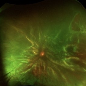
 Multiple Tear RD
Oct 10 2025 by Angela Rico
67 year-old male with complaint of floaters and veil OD for 2 weeks.
Condition/keywords: horseshoe tear, retinal detachment, Retinal tear
-
 Giant Retinal Tear
Sep 20 2025 by JORGE SOBERANES
A complex case of retina detachment due to giant retinal tear
Photographer: Dr. Jorge Soberanes, Asociación para Evitar la Ceguera en México (APEC), UNAM
Condition/keywords: giant retinal tear, Retina detachment
-
 GRT Detachment of 10+ Clock Hours With Folded Retina
Sep 11 2025 by Luis J Haddock, MD
Fundus photo of giant retinal tear detachment involving 10+ hours of continuous tearing of the retina, visible anterior edge of retina over GRT.
Photographer: Natella Romero, University of Miami, Bascom Palmer Eye Institute
Imaging device: Optos
Condition/keywords: acute retinal detachment, Giant retinal tear
-
 Retinal Tear
Sep 4 2025 by Kimberly Wakester
Optomap RBG of a 55-year-old woman with a retinal tear at 12 with bridging vessel and some fluid. Treatment with prophylaxis laser was recommended. Patient is to continue follow up care post operatively.
Photographer: Kimberly Wakester, COA, OCT-C
Imaging device: Optos California
Condition/keywords: left eye, PVD, Retinal tear
-
 Retinal Tear w/VH
Aug 22 2025 by Virginia Gebhart
69 year old male referred for sudden vision loss. Difficult view secondary to VH. Ultrasound and photos show small break with clotting, heavy amount of layered blood inferior and scattered IRH's. Cryotherapy performed to seal current tear, will give VH time to clear on its own. Pt takes Eliquis twice a day.
Photographer: Virginia Gebhart, Retina Consultants of Carolina
Imaging device: Optos California
Condition/keywords: cryo-retinal tear, horseshoe tear, tear, vitreous hemorrhage
-
 Post Blunt Trauma Posterior Break
Aug 13 2025 by Debarun Sharma
Fundus photograph of a 21 year old male with history of blunt trauma with fist presenting with a large linear posterior break just adjacent to the infero-temporal arcade with choroidal rupture and subretinal bleed passing through fovea. Successful barrage laser of the break can be seen.
Photographer: Debarun Sharma, Sri Sankardeva Nethralaya, Guwahati
Imaging device: Optos
Condition/keywords: blunt trauma, full thickness retinal tear, laser photocoagulation
-
 Horseshoe Retinal Tear
Aug 6 2025 by Korey Starkey
80 year-old patient presented with HSRT without detachment in the left eye and macula-off detachment in the right eye. Scheduled patient for prompt surgical repair OD and same day laser retinopexy OS to reduce risk of retinal detachment.
Photographer: Korey Starkey
Imaging device: Optos
Condition/keywords: color fundus photograph, fundus photography, horseshoe tear, Optos
-
 Suprachoroidal Hemorrhage
Aug 4 2025 by Anjana Mirajkar, MS Ophthalmology
A fundus photograph of a 56 year old female with a 360 degree suprachoroidal hemorrhage with a 360 degree crumpled retina during cataract surgery.
Photographer: Dr. Anjana Mirajkar- HV Desai eye hospital ,Pune
Imaging device: optos
Condition/keywords: giant retinal tear, suprachoroidal hemorrhage
-
 Retinal Detachment Secondary to Large Temporal Tear
Aug 4 2025 by Anjana Mirajkar, MS Ophthalmology
Fundus photograph of a 55 year old male with a retinal detachment with macula off with a large temporal tear.
Photographer: Dr. Anjana Mirajkar- HV desai eye hospital ,Pune
Imaging device: Optos
Condition/keywords: Retinal Detachment, retinal tear
-
 Multiple Retinal Tears
Jul 25 2025 by Virginia Gebhart
60 year old male referred for horseshoe tear of the retina. Scleral depressed exam revealed 7 tears in the same eye. Prophylaxis laser performed to seal all tears.
Photographer: Virginia Gebhart, Retina Consultants of Carolina
Imaging device: Optos California
Condition/keywords: horseshoe tear, multiple retinal tears, operculated tear, Retinal tear
-
 Jaws of Detachment
Jul 25 2025 by Malvika Singh
Fundus photograph of a 50 year old with a giant retinal tear and vitreous hemorrhage.
Photographer: Dr Malvika Singh, Retina Foundation, Ahmedabad, India
Imaging device: Mirante SLO/OCT
Condition/keywords: giant retinal tear
-
 Retinal detachment with Single Break
Jul 18 2025 by Kimberly Wakester
Optomap RGB of a 62-year-old man with a retinal detachment with a single break in the left eye. Patient has a previously treated HSRT in the left eye. Surgery was recommended. Patient is to continue follow up care post operatively.
Photographer: Kimberly Wakester, COA, OCT-C
Imaging device: Optos California
Condition/keywords: RD, retinal tear
-
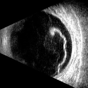 Giant Retinal Tear
Jul 5 2025 by Gustavo Uriel Fonseca Aguirre
This B-mode longitudinal ultrasound scan reveals a giant retinal tear, demonstrating a circumferential retinal flap with rolled edges extending over M-X to M-I. The vitreous shows diffuse hemorrhage and anterior-posterior traction strands inserting at the tear margins. The remaining retina appears attached without subretinal fluid.
Photographer: Gustavo U. Fonseca Aguirre, Hospital Conde de Valenciana, Ciudad de México
Condition/keywords: giant retinal tear
-
 Retinal Detachment with Giant Retinal Tear
May 14 2025 by Kimberly Wakester
Optomap RGB of an 66-year-old man with a retinal detachment with a giant retinal tear in the right eye. Surgery was recommended. Patient is to continue follow up care post operatively. Also noted in the image is a vitreous opacity that was caught at the right moment and appears to look like a smiley face.
Photographer: Kimberly Wakester, COA, OCT-C
Imaging device: Optos California
Condition/keywords: giant retinal tear, RD
-
 Giant Retinal Tear with Multiple Retinal Breaks
Apr 21 2025 by Hrishikesh Naik, MS
A 28 year old high myope with retinal detachment associated with a supero-temporal giant retinal tear in addition to multiple peripheral horseshoe tears and an additional supero-nasal retinal tear.
Photographer: Hrishikesh Naik
Imaging device: Optos Daytona
Condition/keywords: giant retinal tear, High Myopia, horseshoe tear, retinal break, retinal detachment
-
 Blister Retinal Detachment Superotemporal with a Flap Tear
Apr 10 2025 by Daniela Bogenschutz
Autofluorescence of a 70-year-old male with a superotemporal retinal detachment prior to having an OCT with unusual findings. Patient states symptoms were "starburst" in his vision in the location of the retinal detachment with the retinal tear. Surgery was scheduled immediately to avoid further progression.
Photographer: Daniela Bogenschutz, OSC; Retina Consultants of the Carolinas, PA
Condition/keywords: Retinal Detachment, retinal detachment with single break
-
 Superior Rhegmatogenous Retinal Detachment (RRD) in the Right Eye, With a Retinal Tear Located Between the 1 and 2 O'clock Positions
Apr 4 2025 by Cesar Orlando Oviedo Vera
A 45-year-old male patient presented with a sudden onset of decreased visual acuity in the right eye, with a 24-hour progression. Upon examination, Image 1 revealed a superior rhegmatogenous retinal detachment in the right eye, with a retinal tear located between the 1 and 2 o'clock positions. Image 2: Pneumatic retinopexy by intravitreal injection of Sulfur Hexafluoride gas (SF6) at the time of diagnosis with subsequent application of 532 nm laser around the retinal tear.
Photographer: Cesar Orlando Oviedo Vera, Hospital Militar de Especialidades Oftalmológicas
Imaging device: Optos
Condition/keywords: Pneumatic Retinopexy, Retinal tear, Rhegmatogenous retinal detachment, SF6, Superior rhegmatogenous retinal detachment
-
 Pneumatic Retinopexy by Intravitreal Injection of Sulfur Hexafluoride Gas (SF6) at the Time of Diagnosis With Subsequent Application of 532 Nm Laser Around the Retinal Tear
Apr 4 2025 by Cesar Orlando Oviedo Vera
A 45-year-old male patient presented with a sudden onset of decreased visual acuity in the right eye, with a 24-hour progression. Upon examination, Image 1 revealed a superior rhegmatogenous retinal detachment in the right eye, with a retinal tear located between the 1 and 2 o'clock positions. Image 2: Pneumatic retinopexy by intravitreal injection of Sulfur Hexafluoride gas (SF6) at the time of diagnosis with subsequent application of 532 nm laser around the retinal tear.
Photographer: Cesar Orlando Oviedo Vera, Hospital Militar de Especialidades Oftalmológicas
Imaging device: Optos
Condition/keywords: Pneumatic Retinopexy, Retinal tear, Rhegmatogenous retinal detachment, SF6, Superior rhegmatogenous retinal detachment
-
 Retinal Detachment with Retinal Tear
Mar 31 2025 by Kimberly Wakester
Optomap RGB of an 48-year-old woman with a retinal detachment with retinal tear in the left eye. Surgery was recommended. Patient is to continue follow up care post operatively.
Photographer: Kimberly Wakester, COA, OCT-C
Imaging device: Optos California
Condition/keywords: Retinal Detachment, retinal tear
-
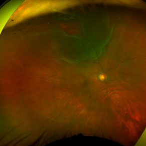
 Rhegmatogenous Retinal Detachment with Gd C PVR Changes
Mar 28 2025 by Shrishti mishra
Fundus photograph of a 58 year old male who had undergone a pneumatic retinopexy elsewhere presented to us with a total retinal detachment with a retinal tear in the superotemporal quadrant and grade c pvr changes.
Photographer: Mrs Vinutha
Imaging device: Optos nikon
Condition/keywords: proliferative vitreoretinopathy (PVR), retinal tear with detachment, rhegmatogenous retinal detachment
-
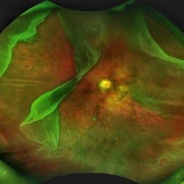
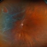
 Rhegmatogenous Retinal Detachment
Mar 24 2025 by DR APOORVA JADHAV, MBBS , DNB
This is a color fundus photograph showing rhegmatogenous retinal detachment with posterior pole retinal tear with macula off.
Condition/keywords: retinal detachment of the macula
-
 Inadvert Globe Perfuration After Peribulbar Block
Mar 13 2025 by Bruno B Ribeiro
Fundus photograph of a 74-year-old woman who underwent pars plana vitrectomy OS due to rhegmatogenous retinal detachment. A horseshoe retinal tear can be seen at 5h. Intraoperative evaluation revealed a chorioretinal scar with the shape of the needle track at the same location. Despite rare, globe perfuration after peri or retrobulbar block may happen, even by the most experienced anesthesiologist.
Photographer: Bruno Barbosa Ribeiro, Angelina Meireles
Imaging device: Optos California
Condition/keywords: retinal detachment
 Loading…