Search results (15 results)
-
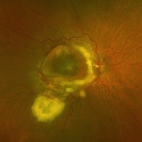 DISCIFORM SCAR AND RETINAL PIGMENT EPITHELIUM (RPE) DETACHMENT IN A CASE OF IDIOPATHIC POLYPOIDAL CHOROIDAL VASCULOPATHY (IPCV)
Oct 21 2023 by Aditya S Kelkar, MS, FRCS, FASRS,FRCOphth
Right eye fundus photograph of a 83 year old female demonstrating Disciform Scar And Retinal Pigment Epithelium (RPE) Detachment In A Case Of Idiopathic Polypoidal Choroidal Vasculopathy (IPCV).
Photographer: DR APURVA MUKADAM
Imaging device: OPTOS DAYTONA
Condition/keywords: disciform scar
-
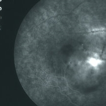 CSR with large RPED
Mar 26 2019 by Gary R. Cook, MD, FACS
Late-phase FA frame showing mild pooling of dye beneath a large RPED inferonasal to the optic disc, blocked fluorescence from the pigment figures (black lines), and late dye leakage from the RPED.
Imaging device: Topcon VT-50
Condition/keywords: central serous retinopathy (CSR), FA late phase, fluorescein angiogram (FA), retinal pigment epithelium (RPE) detachment
-
 CSR with large RPED
Mar 26 2019 by Gary R. Cook, MD, FACS
Mid-phase FA showing large RPED inferonasal to optic disc with overlying cruciate pigment figures (black lines) and neurosensory macular detachment OD.
Imaging device: Topcon VT-50
Condition/keywords: central serous retinopathy (CSR), neurosensory detachment of retina, retinal pigment epithelium (RPE) detachment
-
 CSR with RPED
Mar 26 2019 by Gary R. Cook, MD, FACS
White male with acute CSR with a RPED beneath the NSRD OD.
Imaging device: Topcon VT-50
Condition/keywords: central serous retinopathy (CSR), retinal pigment epithelium (RPE) detachment
-
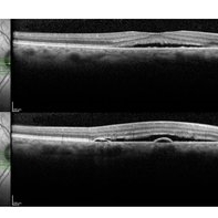 Central Serous Chorioretinopathy-OCT
Jun 22 2018 by Mitzy E Torres Soriano, MD
OCT showing typical subfoveal neurosensory detachment and PEDs in CSR.
Condition/keywords: central serous chorioretinopathy (CSCR), central serous retinopathy (CSR), neurosensory detachment of retina, optical coherence tomography (OCT), retinal pigment epithelium (RPE) detachment
-
 RPE Detachment
Apr 21 2014 by Xiaoxin Li, MD PhD
Multispectral digital ophthalmoscope of a 58-year-old man with a RPE detachment. (810 nm)
Photographer: Xiaoxin Li, Xinxin Zhang
Imaging device: Multispectral digital ophthalmoscope
Condition/keywords: retinal pigment epithelium (RPE) detachment
-
 RPE Detachment
Apr 21 2014 by Xiaoxin Li, MD PhD
Multispectral digital ophthalmoscope of a 58-year-old man with a RPE detachment. (780 nm)
Photographer: Xiaoxin Li, Xinxin Zhang
Imaging device: Multispectral digital ophthalmoscope
Condition/keywords: retinal pigment epithelium (RPE) detachment
-
 RPE Detachment
Apr 21 2014 by Xiaoxin Li, MD PhD
Multispectral digital ophthalmoscope of a 58-year-old man with a RPE detachment. (660 nm)
Photographer: Xiaoxin Li, Xinxin Zhang
Imaging device: Multispectral digital ophthalmoscope
Condition/keywords: retinal pigment epithelium (RPE) detachment
-
 RPE Detachment
Apr 21 2014 by Xiaoxin Li, MD PhD
Multispectral digital ophthalmoscope of a 58-year-old man with a RPE detachment. (580 nm)
Photographer: Xiaoxin Li, Xinxin Zhang
Imaging device: Multispectral digital ophthalmoscope
Condition/keywords: retinal pigment epithelium (RPE) detachment
-
 RPE Detachment
Apr 21 2014 by Xiaoxin Li, MD PhD
Multispectral digital ophthalmoscope of a 58-year-old man with a RPE detachment. (red-green)
Photographer: Xiaoxin Li, Xinxin Zhang
Imaging device: Multispectral digital ophthalmoscope
Condition/keywords: retinal pigment epithelium (RPE) detachment
-
 EDI OCT Detachment With No Subretinal Fluid
Jun 29 2013 by Jason S. Calhoun
A 38-year-old male came in with blurred vision in the left eye. VA is 20/30. Notice a defect inferior of his central vision. Did an fluorescien angiogram to determine an RPE with no subretinal fluid. Also OCT confirms. Patient was injected with Avastin.
Photographer: Jason S. Calhoun, Mayo Clinic Jacksonville, Florida
Imaging device: TOPCON TRC 50-EX/CIRRUS HD OCT
Condition/keywords: central serous retinopathy (CSR), retinal pigment epithelium (RPE) detachment
-
---thumb.JPG/image-square;max$300,300.ImageHandler) Retinal Pigment Epithelial Detachment With No Subretinal Fluid
Retinal Pigment Epithelial Detachment With No Subretinal Fluid
Jun 29 2013 by Jason S. Calhoun
A 38-year-old male who comes in with blurred vision in the left eye. VA is 20/30. Notices a defect inferior of his central vision. Did an fluorescein angiogram to determine an RPE with no sub retinal fluid. Also OCT confirms. Patient was injected with Avastin.
Photographer: Jason S. Calhoun, Mayo Clinic Jacksonville, Florida
Imaging device: TOPCON TRC 50-EX
Condition/keywords: central serous retinopathy (CSR), retinal pigment epithelium (RPE) detachment
-
---thumb.JPG/image-square;max$300,300.ImageHandler) Retinal Pigment Epithelial Detachment With No Subretinal Fluid
Retinal Pigment Epithelial Detachment With No Subretinal Fluid
Jun 29 2013 by Jason S. Calhoun
A 38-year-old male who comes in with blurred vision in the left eye. VA is 20/30. Notices a defect inferior of his central vision. Did an fluorescein angiogram to determine an RPE with no subretinal fluid. Also OCT confirms. Patient was injected with Avastin.
Photographer: Jason S. Calhoun, Mayo Clinic Jacksonville, Florida
Imaging device: TOPCON TRC 50-EX
Condition/keywords: central serous retinopathy (CSR), retinal pigment epithelium (RPE) detachment
-
---thumb.JPG/image-square;max$300,300.ImageHandler) Retinal Pigment Epithelial Detachment With No Subretinal Fluid
Retinal Pigment Epithelial Detachment With No Subretinal Fluid
Jun 29 2013 by Jason S. Calhoun
A 38-year-old male who comes in with blurred vision in the left eye. VA is 20/30. Noticed a defect inferior of his central vision. Did an fluorescein angiogram to determine an RPE with no sub retinal fluid. Also OCT confirms. Patient was injected with Avastin.
Photographer: Jason S. Calhoun, Mayo Clinic Jacksonville, Florida
Imaging device: TOPCON TRC 50-EX
Condition/keywords: central serous retinopathy (CSR), retinal pigment epithelium (RPE) detachment
-
 Retinal Pigment Epithelial Detachment With No Subretinal Fluid
Jun 29 2013 by Jason S. Calhoun
A 38-year-old male who came in with blurred vision in the left eye. VA is 20/30. Noticed a defect inferior of his central vision. Did an fluorescein angiogram to determine an RPE with no subretinal fluid. Also OCT confirms. Patient was injected with Avastin.
Photographer: Jason S. Calhoun, Mayo Clinic Jacksonville, Florida
Imaging device: TOPCON TRC 50-EX
Condition/keywords: central serous retinopathy (CSR), retinal pigment epithelium (RPE) detachment
 Loading…