Search results (46 results)
-
 Retinal Artery Macroaneurysm With Macular Edema
Sep 12 2025 by Tejaswita Verma
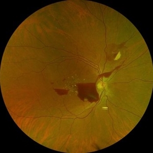
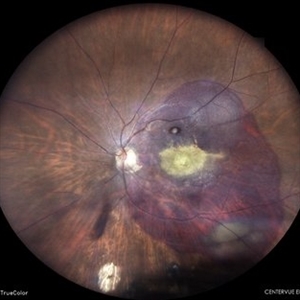
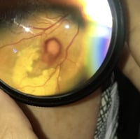
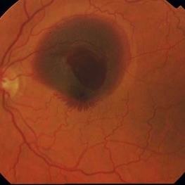
Fundus photo of a 73 year old hypertensive female with 6/18 vision, presenting with RAM ,with surrounding hard exudates and macular edema. She was advised focal laser, anti VEGF injection.
Photographer: DR. TEJASWITA VERMA
Imaging device: MIRANTE
Condition/keywords: RAM, retinal arterial macroaneurysm
-
 Hemorrhagic Retinal Arterial Macroaneurysm
Jul 4 2025 by Julian Navarro Saucedo
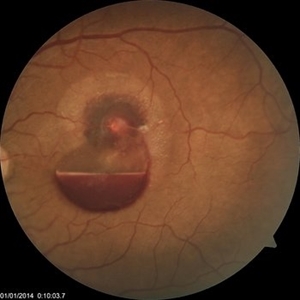
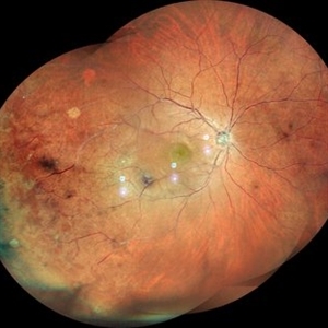
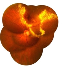
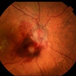
Fundus photograph of a 52-year-old woman with sudden vision loss showing a hemorrhagic retinal arterial macroaneurysm.
Photographer: Julian Navarro, Tecnologico de Monterrey, Escuela de Medician y Ciencias de la Salud.
Imaging device: VISUCAM 524 ZEISS
Condition/keywords: macroaneurysm
-
 The Pouring RAM
Mar 25 2025 by Shrishti mishra
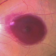
A 63 year old male with RAM lesion in the right eye associated with multilayered hemorrhage.
Imaging device: Optos nikon
Condition/keywords: FFA, retinal arterial macroaneurysm, subhyaloid hemorrhage
-
 Retinal Artery Macro-aneurysms
Jul 19 2024 by Anjana Mirajkar, MS Ophthalmology
An intra operative still of LE showing a retinal artery macro aneurysm causing a sub hylaoid and sub ILM hemorrhage.
Photographer: Dr. Anjana Mirajkar -Retina Foundation, Ahmedabad
Imaging device: Mirante-Nidek
Condition/keywords: retinal arterial macroaneurysm, sub hyaloid hemorrhage, sub internal limiting membrane haemorrhage
-
 Retinal Arterial Macroaneurysm
Jun 5 2024 by Akansha Sharma
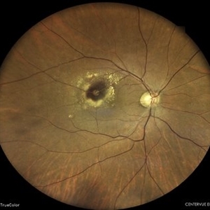
Color fundus photograph of a 61 year old hypertensive male with retinal arterial macroaneurysm.
Photographer: Dr. Akansha Sharma, Bharati Eye Hospital
Condition/keywords: optic disc pallor, RAM
-
 Retinal Arterial Macroaneurysm
Apr 9 2024 by Akansha Sharma
Color fundus photograph of a 68 year old female patient with retinal arterial macroaneurysm with subretinal bleed.
Photographer: Dr. Akansha Sharma, Bharati Eye Hospital
Condition/keywords: macroaneurysm, subretinal hemorrhage
-
 Choroidal Mass
Mar 4 2024 by ANKIT JAIN
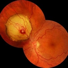
RE color photo montage of right eye of 48 year old with sub retinal hemorrhage with sub retinal fluid at level of fovea.
Photographer: Dr Ankit Jain
Imaging device: MIRANTE
Condition/keywords: macroaneurysm, retinal arterial macroaneurysm
-
 Macroaneurysms
Jan 28 2024 by Anjana Mirajkar, MS Ophthalmology
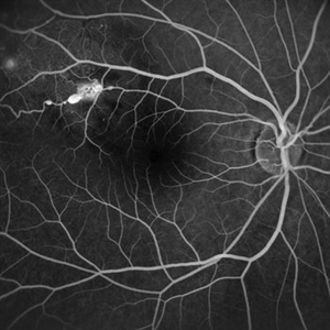
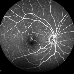
Fundus fluorescein angiography image (Late phase) in a 20 year old female showing leakage in a case of retinal artery macro-aneurysms.
Photographer: Dr. Anjana Mirajkar -Retina Foundation, Ahmedabad
Imaging device: Mirante-Nidek
Condition/keywords: retinal arterial macroaneurysm
-
 Macroaneurysms
Jan 28 2024 by Anjana Mirajkar, MS Ophthalmology
Fundus image of post focal laser in a 20 year old female in a case of retinal arterial macroaneurysm
Photographer: Dr. Anjana Mirajkar -Retina Foundation, Ahmedabad
Imaging device: Mirante-Nidek
Condition/keywords: macroarterial aneurysm
-
 Ruptured retinal artery macroaneurysm
Sep 6 2023 by PRATIK SHENOY, MBBS, DNB, FVRS
A 66-year-old female presented with a ruptured retinal artery macroaneurysm and a visual acuity of finger counting close to face. The multi layered hemorrhage receded on its own inferiorly with an improvement of visual acuity. However, the patient developed a breakthrough bleed with vitreous hemorrhage three weeks later with a drop in visual acuity to hand movements. She underwent pars plana vitrectomy for the same with an improvement in visual acuity to 6/9.
Photographer: Gaurav Kamble, Isha Netralaya
Imaging device: Optos
Condition/keywords: OCT, Optos, pars plana vitrectomy (PPV), retinal arterial macroaneurysm, vitreous hemorrhage
-
 RETINAL ARTERIAL MACROANEURYSM
Jun 6 2023 by Akansha Sharma
COLOUR FUNDUS PHOTOGRAPH OF A 60 YEAR OLD HYPERTENSIVE MALE WITH RETINAL ARTERIAL MACROANEURYSM LEADING TO SUBRETINAL BLEED
Photographer: Dr. Urmil Shah, Dr. Denish Patel, Dr. Akansha Sharma, BHARATI EYE CLINIC, AHMEDABAD, GUJARAT
Condition/keywords: retinal arterial macroaneurysm
-
 Retinal Arterial Macroaneurysm
Apr 8 2023 by Yousef A Fouad, MD, FRCS (Glasg.)
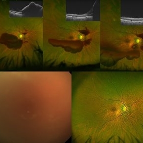
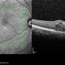
Multimodal imaging of a retinal arterial macroaneurysm in the right eye of a 73-year-old male patient with uncontrolled hypertension. Fundus photography shows hemorrhage surrounding an arterial branch of the upper temporal arcade. Optical coherence tomography (OCT) through the lesion shows inner retinal hyperreflectivity with back shadowing, and adjacent cystoid macular edema in the outer retina. En face OCT centered on the lesion delineates the fusiform dilatation of the affected vessel, and OCT angiography confirms the presence of blood flow within the aneurysmal dilatation.
Photographer: Yousef Fouad, Ain Shams University, Egypt
Condition/keywords: arteriolar macroaneurysm, enface imaging, macroaneurysm, macroarterial aneurysm, OCT Angiography, OCTA
-
 Retinal Arterial Macroaneurysm
Jun 24 2021 by Gabriel Costa Andrade, PhD
Fundus photograph of an 67-year-old woman with a retinal arterial macroaneurysm in macula area with associated exudation.
Photographer: Gabriel Andrade
Condition/keywords: macroaneurysm
-
 Sub ILM Bleed in a Case of Retinal Arterial Macroaneurysm
Mar 11 2021 by Navneet Mehrotra, DNB
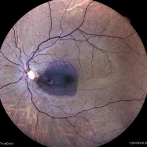
Fundus photograph of a 42-year-old female, who presented with sudden diminution of vision since morning.
Photographer: Navneet Mehrotra
Imaging device: Nidek RS 330
Condition/keywords: retinal arterial macroaneurysm
-
 Ruptured Retinal Arterial Macroaneurysm
May 21 2020 by Elias Khalili Pour, MD
Ruptured Retinal Arterial Macroaneurysm in a patient with history of hypertension
Condition/keywords: cavernous hemangioma of the retina
-
 RAM With Garland of Hard Exudate
Mar 3 2020 by KRISHNENDU NANDI, MS
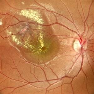
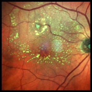
Fundus Photo of left eye of 75-year-old female with retinal artery macroaneurysm at superior quadrant with garland like hard exudates.
Photographer: KRISHNENDU NANDI
Imaging device: Topcon
Condition/keywords: hard exudates, retinal arterial macroaneurysm
-
 Retinal Artery Macroaneurysm
Sep 29 2019 by Haider Ali
65-year-old male with progressively decreasing vision in his left eye for last three months.
Photographer: Dr Haider Ali Chaudhry, Madinah Teaching Hospital
Imaging device: Retinal Artery Macroaneurysm
Condition/keywords: arteriolar macroaneurysm, hypertensive retinopathy, retinal arterial macroaneurysm
-
 Retinal Arterial Macroaneurism
Sep 17 2019 by Zachary M Bodnar, MD
Retinal arterial macroaneurism.
Condition/keywords: retinal arterial macroaneurysm
-
 Aneurysm Early
Sep 17 2019 by Zachary M Bodnar, MD
Aneurysm early
Condition/keywords: retinal arterial macroaneurysm
-
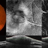
 Aneurysm OCT
Sep 17 2019 by Zachary M Bodnar, MD
Aneurysm OCT
Condition/keywords: optical coherence tomography (OCT), retinal arterial macroaneurysm
-
 Ruptured Retinal Arterial Macroaneurysm
Jun 14 2019 by Niloofar Piri, MD
Fundus photograph of the left eye demonstrating classic hemorrhages in all three layers of retina (subretinal, intra-retinal (radiating pattern around fovea), and sub-ILM in the center), secondary to ruptured retinal arterial macroaneurysm
Condition/keywords: retinal arterial macroaneurysm, ruptured macroaneurysm
-
 Slide 9-19
Feb 26 2019 by Lancaster Course in Ophthalmology
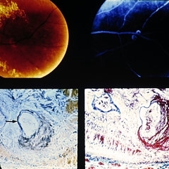
Retinal arterial macroaneurysm. A ring of retinal exudate partially surrounds the macroaneurysm (upper left), which is more clearly delineated by fluorescein (upper right). The retinal arteriole is greatly dilated, and the stain for elastic tissue shows a localized area of disruption and loss of the internal elastic membrane (arrow). The surrounding retina is thickened by edema and some hemorrhage. The ectatic area of the vessel wall is greatly thickened by the accumulation of a laminated fibrinous material. (Courtesy of Alan Friedman, M.D.)
Condition/keywords: retinal arterial macroaneurysm, retinal exudates
-
 Retinal Arterial Macroaneurysm
Sep 18 2018 by Somnath Chakraborty, MD
Left eye fundus photo of a 72-year-old hypertensive, female with a hemorrhagic retinal arterial macroaneurysm with sub-retinal blood.
Photographer: Pulok Chandra Roy, Retina Institute of Bengal
Condition/keywords: retinal arterial macroaneurysm
-
 Retinal Arterial Macroaneurysm
May 3 2018 by Alexandr Stepanov
Retinal arterial macroaneurysm.
Photographer: Alexandr Stepanov MD, PhD, FEBO, Faculty Hospital Hradec Kralove, Czech Republic
Condition/keywords: retinal arterial macroaneurysm
-
 Macroaneurysm
Feb 6 2018 by Bastián Schmidt Arias
Fundus photograph of an 90-year-old woman with a retinal arterial macroaneurysm.
Photographer: Bastian Schmidt
Imaging device: TRC-50DX - Topcon
Condition/keywords: fundus photograph, macroaneurysm
 Loading…