Search results (493 results)
-
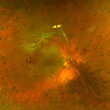
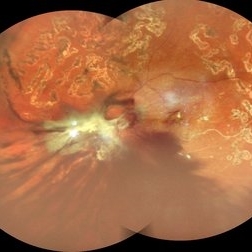 Table Top Tractional Retinal Detachment With Vitreous Hemorrhage in a Case of Proliferative Diabetic Retinopathy
Sep 12 2025 by Akansha Sharma
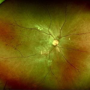
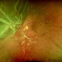
Color fundus photograph of a 56 year old male with table top tractional retinal detachment with vitreous hemorrhage in a case of proliferative diabetic retinopathy.
Photographer: DR. AKANSHA SHARMA
Condition/keywords: pan-retinal photocoagulation (PRP), PDR, proliferative diabetic retinopathy (PDR), PRP, TABLE TOP TRD, tractional retinal detachment, TRD, VH, vitreous hemorrhage
-
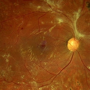 Macular Hole Due to Proliferative Diabetic Retinopathy
Aug 13 2025 by Ricardo Leitão Guerra
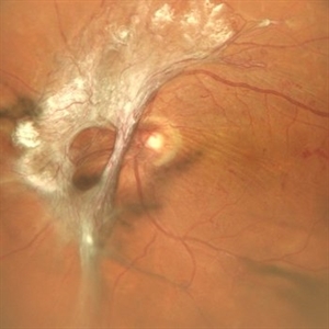
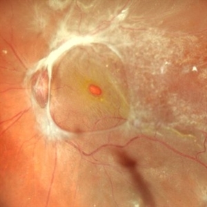
A macular hole formation after anti-VEGF injection prior to vitrectomy for tractional retinal detachment in a patient presenting proliferative diabetic retinopathy.
Photographer: Ricardo Leitão Guerra
Imaging device: ZEISS CLARUS 700
Condition/keywords: macular hole, proliferative diabetic retinopathy (PDR)
-
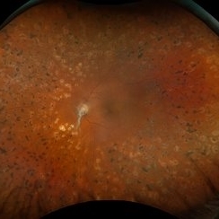 Proliferative Diabetic Retinopathy
Aug 11 2025 by Marin Shehata
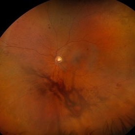
Fundus photograph of a 63 year-old male with diabetic retinopathy has been treated with PRP.
Photographer: Marin Shehata, Retina Consultants of Carolina
Imaging device: Optos California
Condition/keywords: proliferative diabetic retinopathy (PDR), PRP
-
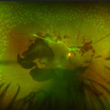 Unstable PDR s/p Laser
Aug 4 2025 by Anjana Mirajkar, MS Ophthalmology
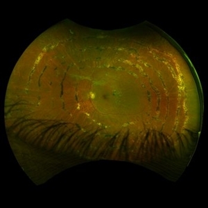
Fundus photograph of a 60 year old male with an unstable PDR showing traction at the posterior pole with sub hyaloid hemorrhage. Peripheral PRP marks can be seen.
Photographer: Dr. Anjana Mirajkar- HV Desai eye hospital ,Pune
Imaging device: Optos
Condition/keywords: pan-retinal photocoagulation (PRP), proliferative diabetic retinopathy (PDR), subhyaloid hemorrhage, tractional retinal detachment
-
 Shooting Stars
Jul 9 2025 by Majda Hadziahmetovic, MD
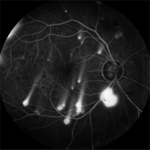
Fluorescein angiography image demonstrating multiple areas of neovascularization in a middle-aged male patient with long-standing diabetes.
Condition/keywords: proliferative diabetic retinopathy (PDR)
-
 Proliferative Diabetic Retinopathy
Jul 9 2025 by Jeffrey Barker
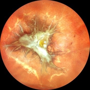
57 year old male presents with Proliferative Diabetic Retinopathy and Tractional Retina detachment.
Photographer: Jeffrey P. Barker, B.S. Retina Vitreous Surgeons of CNY
Condition/keywords: Diabetes, proliferative diabetic retinopathy (PDR), Traction retinal detachment
-
 Traction in Proliferative Diabetic Retinopathy
Jun 9 2025 by Malvika Singh
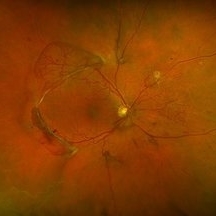
Fundus photograph of a 44 year old with uncontrolled diabetes showing fibrovascular proliferation and traction with details of disc and macula obscured with sclerosed vessels in the periphery.
Photographer: Dr Malvika Singh, Retina Foundation, Ahmedabad, India
Imaging device: Mirante SLO/OCT
Condition/keywords: proliferative diabetic retinopathy (PDR), TRACTION
-
 Neovascularization of the Disc
Jun 3 2025 by Scott D Walter, MD, MSc, FASRS
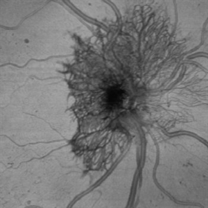
Near-infrared (NIR) en face OCT image showing neovascularization of the disc (NVD) in a patient with type II diabetes mellitus, complicated by proliferative diabetic retinopathy (PDR).
Imaging device: Heidelberg Spectralis
Condition/keywords: Diabetes, Heidelburg Spectralis, microaneurysms, Neovascularisation at the Disc (NVD), NEOVASCULARISATION OF DISC, OCT EN FACE, proliferative diabetic retinopathy (PDR)
-
 The Dread of the Crimson Red
Jun 2 2025 by Thirumalesh Mochi Basavaraj, MD
Fundus photograph of a 64 year man post laser depicting a regressed NVD in the superior aspect and a Persistent Neo vascularization in the inferior aspect
Photographer: Vivek
Condition/keywords: Neovascularisation at the Disc (NVD), proliferative diabetic retinopathy (PDR)
-
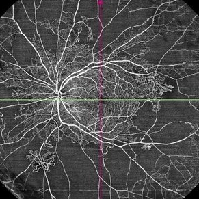 Proliferative Diabetic Retinopathy
May 29 2025 by KANWALJEET HARJOT MADAN, M.S. (Ophthalmology); FAICO (Vitreous - Retina)
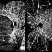
This is widefield optic coherence tomography angiography (WF-OCTA) picture of LE of a diabetic patient. This patient had Proliferative Diabetic Retinopathy and depicts large areas of capillary non perfusion with neovascularization elsewhere.
Photographer: Dr. Kanwaljeet Harjot Madan, Thind Eye Hospital, Jalandhar City (Punjab) INDIA.
Imaging device: Widefield Optic Coherence Tomography Angiography (WF-OCTA).
Condition/keywords: OCTA, proliferative diabetic retinopathy (PDR), ultra-wide field imaging
-
 High Risk Proliferative Diabetic Retinopathy with Sub-hyaloid Hemorrhage
May 13 2025 by Anupama Kiran Kumar
This image shows a case of high risk proliferative diabetic retinopathy. The retina is unlasered with a taut posterior hyaloid and a sub-hyaloid hemorrhage at the macula and along the arcades ,sparing the fovea.
Photographer: Mr Pratap
Imaging device: Mirante SLO/OCT (Nidek Co., Gamagori, Japan)
Condition/keywords: Diabetes, Diabetic Retinopathy, proliferative diabetic retinopathy (PDR), subhyaloid hemorrhage
-
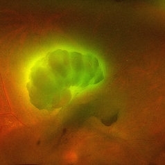 Aurora Borealis in Retina
Apr 25 2025 by Poornachandra B, MS, FVRS
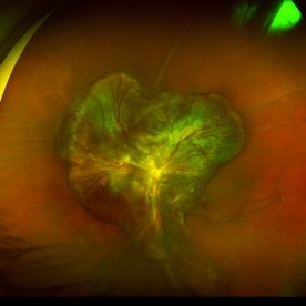
Fundus picture of 54 year old male with proliferative diabetic retinopathy with fluorescent blood clot in vitreous cavity.
Photographer: Mr Dhikshith
Imaging device: Optos daytona
Condition/keywords: blood, proliferative diabetic retinopathy (PDR)
-
 Bilateral Proliferative Diabetic Retinopathy OU
Feb 21 2025 by Drew Mitchell
OCT-Angiography 8x8 Montage OU. PDR with active NVE OD. 37 year old male with no visual complaints. Vision is 20/20 in both eyes.
Photographer: Drew Mitchell OCT-C
Imaging device: Zeiss Cirrus 5000
Condition/keywords: CIRRUS 5000 ANGIOPLEX, Diabetes, NVE, OCT Angiography, proliferative diabetic retinopathy (PDR)
-
 Vascular maze- Proliferative Diabetic Retinopathy
Feb 7 2025 by Hemanth Murthy, MBBS, MD, FASRS
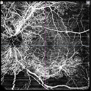
OCTA image left eye. A 32 year male with history of blurring of vision in right eye since 4 months. History of Diabetes and Hypertension since 2 years. Vision 6/36 in right eye and 6/9 in left eye
Photographer: Veda Vyas
Condition/keywords: proliferative diabetic retinopathy (PDR)
-
 Vascular Maze-Proliferative Diabetic Retinopathy
Feb 7 2025 by Hemanth Murthy, MBBS, MD, FASRS
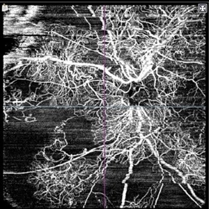
OCTA image right eye-A 32 year male with history of blurring of vision in right eye since 4 months. History of Diabetes and Hypertension since 2 years. Vision 6/36 in right eye and 6/9 in left eye
Photographer: Veda Vyas
Condition/keywords: OCT Angiography, proliferative diabetic retinopathy (PDR)
-
 Vascular Maze-Proliferative Diabetic Retinopathy
Feb 7 2025 by Hemanth Murthy, MBBS, MD, FASRS
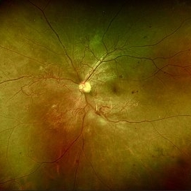
Fundus photo left eye. A 32 year male with history of blurring of vision in right eye since 4 months. History of Diabetes and Hypertension since 2 years. Vision 6/36 in right eye and 6/9 in left eye
Photographer: Veda Vyas
Condition/keywords: proliferative diabetic retinopathy (PDR)
-
 Vascular Maze-Proliferative Diabetic Retinopathy
Feb 7 2025 by Hemanth Murthy, MBBS, MD, FASRS
Fundus photo of right eye. A 32 year male with history of blurring of vision in right eye since 4 months. History of Diabetes and Hypertension since 2 years. Vision 6/36 in right eye and 6/9 in left eye
Photographer: Veda Vyas
Condition/keywords: proliferative diabetic retinopathy (PDR)
-
 PDR with NVD
Dec 5 2024 by Tejaswita Verma
Fundus image of a middle aged male with NVD, multiple dot blot and flame shaped hemorrhages, cotton wool spots, hard exudates at the posterior pole in a case of PDR . Vision was 6/9.
Photographer: DR. TEJASWITA VERMA
Imaging device: MIRANTE
Condition/keywords: NEOVASCULARISATION OF DISC, proliferative diabetic retinopathy (PDR)
-
 PDR/Vitreous Hemorrhage
Nov 20 2024 by Virginia Gebhart
76 year old male with new proliferative diabetic retinopathy. NVD and VH on exam. Pt treated with IVEylea, will consider PRP in the near future.
Photographer: Virginia Gebhart, Retina Consultants of Carolina
Imaging device: Optos California
Condition/keywords: PDR, proliferative diabetic retinopathy (PDR), VH, vitreous hemorrhage
-
 Panretinal Photocoagulation
Nov 7 2024 by Ellen Y Kang
Fundus photograph of a 47-year-old male post-op panretinal photocoagulation for proliferative diabetic retinopathy. Fundus revealed procedure had been performed in a spiral.
Condition/keywords: panretinal photo coagulation, proliferative diabetic retinopathy (PDR)
-
 Proliferative Retinopathy
Nov 4 2024 by Tejaswita Verma
Fundus photograph of a middle aged male with diabetes showing large FVP following NVD.
Photographer: DR. TEJASWITA VERMA
Imaging device: MIRANTE
Condition/keywords: FVPs, neovascularization of the disc (NVD), proliferative diabetic retinopathy (PDR)
-
 Combined Traction Rhegmatogenous Detachment
Oct 17 2024 by Hemanth Murthy, MBBS, MD, FASRS
A 68 year old male presented with a shadow in the left eye since 3 days. He was a known diabetic and hypertensive for 20 years. Vision was 20/40 in right eye and 20/60 in left eye. Fundus examination showed Proliferative diabetic retinopathy in right eye and Proliferative diabetic retinopathy with combined traction rhegmatogenous detachment in left eye.
Photographer: Mr Veda Vyas
Condition/keywords: combined retinal detachment, proliferative diabetic retinopathy (PDR)
-
 Proliferative Diabetic Retinopathy
Oct 13 2024 by Brandon I Fram, MD, BS
28 year-old with florid neovascularization of the disc and extensive nonperfusion imaged with fluorescein angiography
Condition/keywords: florid type PDR, fluorescein angiogram (FA), neovascularization of the disc (NVD), PDR, proliferative diabetic retinopathy (PDR)
-
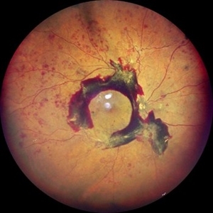
 Combined Retinal Detachment With Macular Hole
Sep 28 2024 by Tejaswita Verma
Fundus image of the LE of a 67 year old diabetic, hypertensive female with CF 3metres vision showing combined RD with FTMH, in a pseudophakic eye. She was lost to follow up status post 2 anti VEGF injections received 8 months back due to typhoid fever.
Photographer: DR. TEJASWITA VERMA
Imaging device: MIRANTE
Condition/keywords: full thickness macular hole, proliferative diabetic retinopathy (PDR), tractional retinal detachment
-
 Retinal Traction Detachment
Aug 31 2024 by Monica Elena Cortizo Brown , MD
A 72 year old man with proliferative diabetic retinopathy with macular traction retinal detachment.
Photographer: Mónica E. Cortizo Brown
Imaging device: Optos
Condition/keywords: macular traction, proliferative diabetic retinopathy (PDR)
 Loading…